The AVPR1A Antibody (CAB8400) is a high-quality antibody developed for reliable detection and analysis of target proteins. This antibody, generated in rabbits, exhibits high specificity and sensitivity towards human samples, making it an ideal choice for Western blot applications.The AVPR1A receptor, also known as the arginine vasopressin receptor 1A, is involved in mediating the effects of the neuropeptide arginine vasopressin (AVP) in the brain. It plays a crucial role in modulating social behaviors such as bonding, aggression, and affiliation, as well as stress-related responses.
This antibody is validated for use in WB, IF/ICC, ELISA applications and has demonstrated reactivity against Human, Mouse, Rat samples.
Product Name:
AVPR1A Antibody
SKU:
CAB8400
Size:
20μL, 100μL
Reactivity:
Human, Mouse, Rat
Conjugate:
Unconjugated
Immunogen:
Recombinant protein (or fragment).This information is considered to be commercially sensitive.
Recommended starting concentration is 1 μg/mL. Please optimize the concentration based on your specific assay requirements.
Synonyms:
V1aR, AVPR1, AVPR V1a, AVPR1A
Positive Sample:
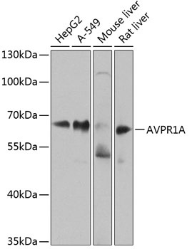
HepG2, A-549, Mouse liver, Rat liver
Cellular Localization:
Cell Membrane, Multi-Pass Membrane Protein.
Calculated MW:
47kDa
Observed MW:
60kDa
The protein encoded by this gene acts as receptor for arginine vasopressin. This receptor belongs to the subfamily of G-protein coupled receptors which includes AVPR1B, V2R and OXT receptors. Its activity is mediated by G proteins which stimulate a phosphatidylinositol-calcium second messenger system. The receptor mediates cell contraction and proliferation, platelet aggregation, release of coagulation factor and glycogenolysis.
Purification Method
Affinity purification
Gene ID
552
RRID
AB_2768529
Buffer Information
Store at -20℃. Avoid freeze / thaw cycles. Buffer: PBS containing 50% glycerol, preserved with proclin300 or sodium azide, pH 7.3.
Western blot analysis of various lysates using AVPR1A Rabbit pAb (CAB8400) at 1:1000 dilution. Secondary antibody: HRP-conjugated Goat anti-Rabbit IgG (H+L) (CABS014) at 1:10000 dilution. Lysates/proteins: 25μg per lane. Blocking buffer: 3% nonfat dry milk in TBST. Detection: ECL Enhanced Kit (AbGn00021). Exposure time: 60s.
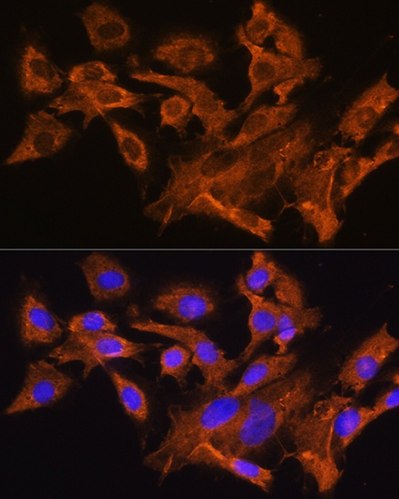
Immunofluorescence analysis of C6 cells using AVPR1A Rabbit pAb (CAB8400) at dilution of 1:100. Secondary antibody: Cy3-conjugated Goat anti-Rabbit IgG (H+L) (CABS007) at 1:500 dilution. Blue: DAPI for nuclear staining.