The BAG1 Antibody (CAB1104) is a high-quality antibody developed for reliable detection and analysis of target proteins. This antibody, derived from rabbits, exhibits high reactivity with human samples and is validated for use in Western blot applications. By binding specifically to the Bag1 protein, this antibody enables accurate detection and analysis in a variety of cell types, making it an ideal choice for studies related to cell biology, cancer research, and apoptosis mechanisms.
This antibody is validated for use in WB, IF/ICC, ELISA applications and has demonstrated reactivity against Human, Mouse samples.
Product Name:
BAG1 Antibody
SKU:
CAB1104
Size:
20μL, 100μL
Reactivity:
Human, Mouse
Conjugate:
Unconjugated
Immunogen:
Recombinant protein (or fragment).This information is considered to be commercially sensitive.
Recommended starting concentration is 1 μg/mL. Please optimize the concentration based on your specific assay requirements.
Synonyms:
HAP, BAG-1, RAP46, BAG1
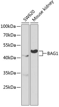
Positive Sample:
SW620, Mouse kidney
Cellular Localization:
Cytoplasm, Cytoplasm, Nucleus.
Calculated MW:
39kDa
Observed MW:
43kDa
The oncogene BCL2 is a membrane protein that blocks a step in a pathway leading to apoptosis or programmed cell death. The protein encoded by this gene binds to BCL2 and is referred to as BCL2-associated athanogene. It enhances the anti-apoptotic effects of BCL2 and represents a link between growth factor receptors and anti-apoptotic mechanisms. Multiple protein isoforms are encoded by this mRNA through the use of a non-AUG (CUG) initiation codon, and three alternative downstream AUG initiation codons. A related pseudogene has been defined on chromosome X.
Purification Method
Affinity purification
Gene ID
573
RRID
AB_2758381
Buffer Information
Store at -20℃. Avoid freeze / thaw cycles. Buffer: PBS containing 50% glycerol, preserved with proclin300 or sodium azide, pH 7.3.
Western blot analysis of various lysates using BAG1 Rabbit pAb (CAB1104) at 1:1000 dilution. Secondary antibody: HRP-conjugated Goat anti-Rabbit IgG (H+L) (CABS014) at 1:10000 dilution. Lysates/proteins: 25μg per lane. Blocking buffer: 3% nonfat dry milk in TBST.
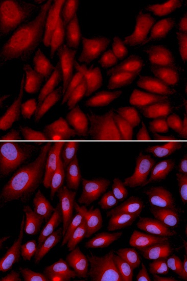
Immunofluorescence analysis of U2OS cells using BAG1 Rabbit pAb (CAB1104). Secondary antibody: Cy3-conjugated Goat anti-Rabbit IgG (H+L) (CABS007) at 1:500 dilution. Blue: DAPI for nuclear staining.