The BAG3 Antibody (CAB14826) is a high-quality antibody developed for reliable detection and analysis of target proteins. This antibody, generated in rabbits, is specifically designed for detecting and analyzing BAG3 in a variety of samples, with a high reactivity towards human samples. Validated for use in Western blot applications, this antibody binds to the BAG3 protein, allowing for accurate and reliable detection in different cell types.BAG3 is known for its role in regulating cell survival and stress responses, making it a key target for research in areas such as cancer, neurodegenerative diseases, and cardiovascular disorders.
This antibody is validated for use in WB, IHC-P, ELISA applications and has demonstrated reactivity against Human, Mouse, Rat samples.
Product Name:
BAG3 Antibody
SKU:
CAB14826
Size:
20μL, 100μL
Reactivity:
Human, Mouse, Rat
Conjugate:
Unconjugated
Immunogen:
Recombinant protein (or fragment).This information is considered to be commercially sensitive.
Recommended starting concentration is 1 μg/mL. Please optimize the concentration based on your specific assay requirements.
Synonyms:
BIS, MFM6, BAG-3, CAIR-1, BAG3
Positive Sample:
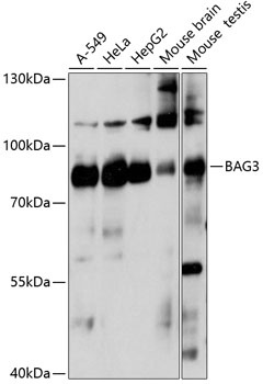
A-549, HeLa, HepG2, Mouse brain, Mouse testis
Cellular Localization:
Cytoplasm, Cytosol, Nucleus, Z Disc.
Calculated MW:
62kDa
Observed MW:
80kDa
BAG proteins compete with Hip for binding to the Hsc70/Hsp70 ATPase domain and promote substrate release. All the BAG proteins have an approximately 45-amino acid BAG domain near the C terminus but differ markedly in their N-terminal regions. The protein encoded by this gene contains a WW domain in the N-terminal region and a BAG domain in the C-terminal region. The BAG domains of BAG1, BAG2, and BAG3 interact specifically with the Hsc70 ATPase domain in vitro and in mammalian cells. All 3 proteins bind with high affinity to the ATPase domain of Hsc70 and inhibit its chaperone activity in a Hip-repressible manner.
Purification Method
Affinity purification
Gene ID
9531
RRID
AB_2761705
Buffer Information
Store at -20℃. Avoid freeze / thaw cycles. Buffer: PBS with 0.01% thimerosal,50% glycerol,pH7.3.
Western blot analysis of various lysates using BAG3 Rabbit pAb (CAB14826) at 1:1000 dilution. Secondary antibody: HRP-conjugated Goat anti-Rabbit IgG (H+L) (CABS014) at 1:10000 dilution. Lysates/proteins: 25μg per lane. Blocking buffer: 3% nonfat dry milk in TBST. Detection: ECL Basic Kit (AbGn00020). Exposure time: 30s.
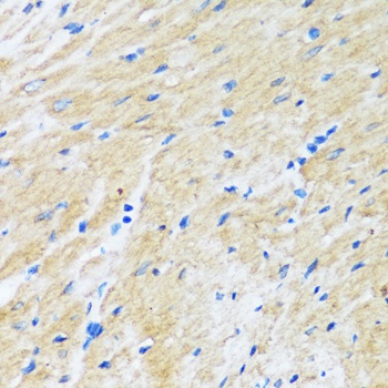
Immunohistochemistry analysis of paraffin-embedded Rat heart using BAG3 Rabbit pAb (CAB14826) at dilution of 1:100 (40x lens). Microwave antigen retrieval performed with 0.01M PBS Buffer (pH 7.2) prior to IHC staining.
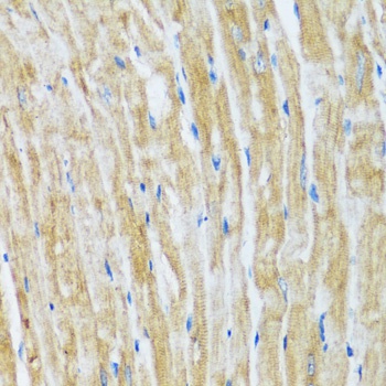
Immunohistochemistry analysis of paraffin-embedded Mouse heart using BAG3 Rabbit pAb (CAB14826) at dilution of 1:100 (40x lens). Microwave antigen retrieval performed with 0.01M PBS Buffer (pH 7.2) prior to IHC staining.