
Understanding the Autophagy Pathway: A Critical Process in Cellular Maintenance
The Autophagy Pathway: Mechanism, Regulation and How to Measure It
Autophagy is the cell’s route for degrading and recycling its own contents — damaged organelles, aggregated protein, intracellular pathogens. The mechanism is well mapped, from ULK1 initiation through to lysosomal degradation, and it sits under a single regulatory switch formed by mTORC1 and AMPK. This guide covers the pathway, its regulation, the selective forms, and the flux measurement problem that makes autophagy easy to assay badly.
Browse autophagy antibodies →Key takeaways
- Autophagy degrades and recycles cellular components through the lysosome, clearing damaged organelles and misfolded protein and releasing nutrients under starvation.
- Three forms exist: macroautophagy (the autophagosome route), microautophagy (direct lysosomal engulfment) and chaperone-mediated autophagy (selective, receptor-driven).
- Regulation converges on ULK1: mTORC1 phosphorylates it at Ser757 to suppress autophagy, while AMPK phosphorylates it at Ser317 and Ser556 to activate it.
- The two ubiquitin-like conjugation systems are ATG12–ATG5–ATG16L1 and the LC3/ATG8 system. LC3-II is the product of the second — LC3 conjugated to phosphatidylethanolamine — not the system itself.
- Much of autophagy is selective, not bulk: mitophagy, xenophagy, aggrephagy, lipophagy and ER-phagy each use dedicated cargo receptors.
- Rising LC3-II does not mean autophagy increased. It can equally mean degradation is blocked. Autophagic flux must be measured with and without a lysosomal inhibitor.
- In cancer autophagy is genuinely dual — restraining transformation early, then sustaining established tumours under metabolic stress.
Antibodies for autophagy research
The panel covers the marker and its flux partner, the nucleation complex, and both arms of the regulatory switch converging on ULK1.
![[KO Validated] LC3B Rabbit Polyclonal Antibody](https://cdn11.bigcommerce.com/s-h68l9z2lnx/products/77236/images/462568/anti-lc3b-antibody-cab11282ko-validated__14740.1631918453.386.513.jpg?c=2)
[KO Validated] LC3B Rabbit Polyclonal Antibody
The core autophagy marker; knockout validation matters here because LC3 antibodies vary widely in specificity.
View product →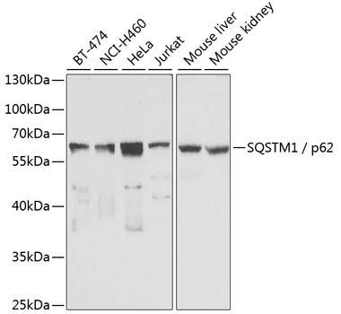
SQSTM1 / p62 Rabbit Polyclonal Antibody
The cargo receptor degraded along with its cargo — read alongside LC3-II to interpret flux.
View product →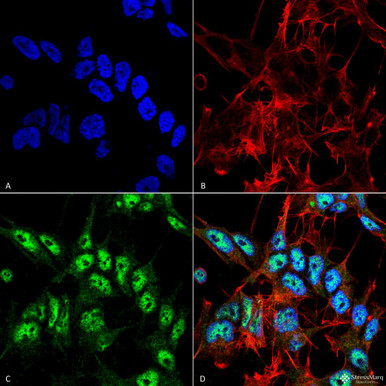
Anti-Beclin 1 Antibody
Part of the class III PI3K nucleation complex, and the node where autophagy meets Bcl-2 signalling.
View product →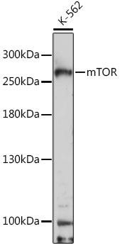
mTOR Rabbit Polyclonal Antibody
The master suppressor — active mTORC1 keeps autophagy off under nutrient-replete conditions.
View product →
Phospho-AMPKalpha (Thr172 / Thr183) Monoclonal Antibody
Thr172 is the activating site; phosphorylation here reports the energy-stress signal that switches autophagy on.
View product →
Anti-Phospho-ULK1 (Ser556) Monoclonal Antibody
The AMPK site on ULK1 — a direct readout of the activating arm at the convergence point.
View product →What autophagy is
Autophagy — from the Greek auto, self, and phagy, eating — is the process by which a cell degrades and recycles its own components through the lysosome. It clears damaged organelles, misfolded and aggregated protein and intracellular pathogens, and under starvation it liberates amino acids and fatty acids to keep the cell alive.
It runs constitutively at a low level as housekeeping, and is upregulated sharply under stress. The mechanism was worked out largely in yeast genetics, and Yoshinori Ohsumi received the 2016 Nobel Prize in Physiology or Medicine for that work.
It is worth distinguishing autophagy from the ubiquitin-proteasome system, the cell’s other major degradation route. The proteasome handles short-lived individual proteins threaded through a narrow barrel; autophagy handles bulk cargo too large for that — whole organelles, protein aggregates, bacteria.
The three types
| Type | Mechanism | Cargo |
|---|---|---|
| Macroautophagy | A double-membraned autophagosome forms around cargo and fuses with the lysosome | Organelles, aggregates, pathogens, bulk cytoplasm |
| Microautophagy | The lysosomal membrane invaginates and engulfs cargo directly | Small cytosolic portions; some organelles |
| Chaperone-mediated autophagy | HSC70 recognises a KFERQ-like motif and delivers the protein through LAMP-2A for unfolding and import | Individual soluble proteins only |
Macroautophagy is what is usually meant by “autophagy” unqualified, and it is what the rest of this guide describes. Chaperone-mediated autophagy is notably different in that cargo is translocated one unfolded protein at a time rather than engulfed in bulk.
The pathway step by step
- Initiation. Nutrient deprivation, hypoxia or oxidative stress activate the ULK1 complex (ULK1 with ATG13, FIP200 and ATG101), which nucleates the isolation membrane.
- Nucleation. The class III PI3K complex — VPS34 with Beclin-1, ATG14L and VPS15 — generates phosphatidylinositol 3-phosphate on the nascent phagophore, recruiting PI3P-binding effectors such as WIPI2 and DFCP1.
- Elongation. Two ubiquitin-like conjugation systems expand the membrane. The first produces the ATG12–ATG5–ATG16L1 complex; the second conjugates LC3 to phosphatidylethanolamine, converting soluble LC3-I into membrane-bound LC3-II, which decorates both faces of the growing phagophore.
- Cargo selection. Receptors including p62/SQSTM1, NBR1 and optineurin bridge ubiquitinated cargo to LC3 through an LIR motif, giving selectivity.
- Closure and fusion. The phagophore seals into a double-membraned autophagosome, which fuses with a lysosome. Fusion requires SNAREs — STX17 and SNAP29 on the autophagosome with VAMP8 on the lysosome — plus tethering by the HOPS complex.
- Degradation and recycling. Lysosomal hydrolases degrade the inner membrane and contents; amino acids, lipids and sugars are exported back to the cytosol, and lysosomes are regenerated by autophagic lysosome reformation.
Two details worth holding onto. LC3-II sits on both membranes, so the inner pool is degraded with the cargo while the outer pool is recycled — which is exactly why LC3-II levels alone are ambiguous. And p62 is itself an autophagy substrate, so it falls when flux is working and accumulates when flux is blocked.
Regulation: mTORC1 and AMPK
The original article described the pathway without saying what controls it, yet the regulation is the part most experiments actually manipulate. Two nutrient sensors act in opposition, and both converge on ULK1.
| Sensor | Activated by | Effect on autophagy |
|---|---|---|
| mTORC1 | Amino acids, growth factors, high energy | Suppresses. Phosphorylates ULK1 at Ser757, preventing AMPK from activating it, and phosphorylates ATG13 and TFEB |
| AMPK | Rising AMP:ATP ratio — energy stress | Activates. Phosphorylates ULK1 at Ser317 and Ser556, and inhibits mTORC1 indirectly via TSC2 and raptor |
Under nutrient-rich conditions mTORC1 is active on the lysosomal surface and autophagy is held off. Starvation inactivates mTORC1 and activates AMPK, and ULK1 is released to initiate. Because the two act on different residues of the same protein, phospho-specific ULK1 antibodies distinguish which arm is driving a response.
A third layer operates transcriptionally: mTORC1 phosphorylation retains TFEB in the cytosol, and on mTORC1 inhibition TFEB enters the nucleus and induces lysosomal and autophagy genes — coordinating the longer-term capacity of the system rather than the immediate response.
Selective autophagy
Autophagy is often described as bulk degradation, but most of it is highly selective, with a dedicated receptor for each cargo type.
| Type | Cargo | Key receptors |
|---|---|---|
| Mitophagy | Damaged mitochondria | PINK1–Parkin ubiquitination with optineurin and NDP52; BNIP3 and NIX in receptor-mediated form |
| Xenophagy | Intracellular bacteria and viruses | p62, NDP52, optineurin, with galectin-8 marking damaged vacuoles |
| Aggrephagy | Protein aggregates | p62 and NBR1 |
| Lipophagy | Lipid droplets | Regulates cellular lipid stores and hepatic steatosis |
| ER-phagy | Endoplasmic reticulum fragments | FAM134B, SEC62, RTN3 |
| Ferritinophagy | Ferritin, releasing iron | NCOA4 — links autophagy to ferroptosis |
Mitophagy is the best characterised and the most clinically resonant: PINK1 and Parkin are both mutated in familial Parkinson’s disease, which connects the general observation that autophagy is impaired in neurodegeneration to a specific molecular mechanism.
Measuring autophagy and the flux problem
This is where autophagy experiments most often go wrong, and it deserves stating plainly.
An increase in LC3-II does not demonstrate that autophagy has increased. LC3-II accumulates either because more autophagosomes are being made, or because they are not being degraded. Those are opposite conclusions from the same blot.
Measuring flux properly
- Compare with and without a lysosomal inhibitor. Run parallel samples with bafilomycin A1 or chloroquine, which block degradation. If flux is active, LC3-II rises further in the inhibited sample; if the difference between the two is unchanged, the apparent increase reflected a block rather than induction.
- Read p62 alongside. p62 is degraded by autophagy, so genuine induction lowers it while a block raises it — moving opposite to LC3-II under induction.
- Report the LC3-II/LC3-I ratio with caution. The two forms transfer and stain differently; LC3-II against a loading control is generally the safer measure.
- Tandem mCherry-GFP-LC3 distinguishes stages by fluorescence: GFP is quenched in the acidic autolysosome while mCherry persists, so autophagosomes appear yellow and autolysosomes red.
- Electron microscopy remains the reference method for identifying genuine double-membraned autophagosomes.
The community standard for these decisions is Klionsky and colleagues’ periodically updated guidelines, listed in the references below.
Autophagy in health and disease
Physiological roles
- Survival under starvation — recycling supplies amino acids and fatty acids when external nutrients are absent.
- Development and differentiation — large-scale organelle clearance, as in erythrocyte and lens fibre maturation.
- Quality control — continuous removal of damaged mitochondria and aggregated protein, particularly important in long-lived post-mitotic cells such as neurons.
- Immunity — xenophagy destroys intracellular pathogens, and autophagy contributes to antigen presentation on MHC class II.
Disease associations
- Cancer — genuinely dual. Early on autophagy is tumour-suppressive: BECN1 is monoallelically deleted in a substantial fraction of breast and ovarian cancers, and loss permits accumulation of damaged mitochondria and genomic instability. In established tumours it becomes supportive, sustaining cells through hypoxia and nutrient limitation, and RAS-driven tumours in particular are autophagy-dependent.
- Neurodegeneration — impaired clearance underlies the aggregates of Alzheimer’s, Parkinson’s and Huntington’s disease, with PINK1 and Parkin mutations giving a direct mitophagy link.
- Infection — several pathogens subvert the pathway. Mycobacterium tuberculosis blocks autophagosome-lysosome fusion, and some viruses use autophagic membranes as replication platforms.
- Metabolic and inflammatory disease — ATG16L1 variants are an established Crohn’s disease risk factor, and lipophagy defects contribute to hepatic steatosis.
Therapeutic implications
Because the direction of benefit depends on context, autophagy is targeted both ways.
- Inhibition in oncology. Chloroquine and hydroxychloroquine block lysosomal acidification and have been trialled alongside chemotherapy in autophagy-dependent tumours. They are blunt tools — lysosomal function is affected broadly, not autophagy specifically.
- Induction in neurodegeneration. Enhancing clearance of aggregated protein is attractive; rapamycin and its analogues induce autophagy through mTORC1 inhibition, though at the cost of immunosuppression and metabolic effects.
- Caloric restriction and exercise both activate AMPK and induce autophagy, which is one proposed mechanism behind their effects on healthspan.
- Biomarkers — LC3-II and p62 in tissue are being explored for stratification, though the flux problem above complicates their interpretation in fixed clinical samples.
This describes research directions rather than clinical guidance.
References
- Yang, Z., & Klionsky, D. J. (2010). Eaten alive: a history of macroautophagy. Nature Cell Biology, 12(9), 814–822.
- Levine, B., & Kroemer, G. (2019). Biological Functions of Autophagy Genes: A Disease Perspective. Cell, 176(1-2), 11–42.
- Mizushima, N., & Komatsu, M. (2011). Autophagy: renovation of cells and tissues. Cell, 147(4), 728–741.
- Klionsky, D. J., et al. (2016). Guidelines for the use and interpretation of assays for monitoring autophagy. Autophagy, 12(1), 1–222.
- Jiang, P., & Mizushima, N. (2014). Autophagy and human diseases. Cell Research, 24(1), 69–79.
- Dikic, I., & Elazar, Z. (2018). Mechanism and medical implications of mammalian autophagy. Nature Reviews Molecular Cell Biology, 19(6), 349–364.
- Levine, B., & Deretic, V. (2007). Unveiling the roles of autophagy in innate and adaptive immunity. Nature Reviews Immunology, 7(10), 767–777.
- White, E. (2012). The role for autophagy in cancer. Journal of Clinical Investigation, 122(1), 42–46.
Choosing antibodies
LC3B and p62 for marker and flux, Beclin-1 for the nucleation complex, and mTOR, phospho-AMPK and phospho-ULK1 for the regulatory switch.
Browse autophagy antibodies →Frequently asked questions
What are the three types of autophagy?
Macroautophagy, in which a double-membraned autophagosome engulfs cargo and fuses with the lysosome; microautophagy, where the lysosome invaginates and takes up cargo directly; and chaperone-mediated autophagy, in which HSC70 delivers individual proteins bearing a KFERQ-like motif through LAMP-2A.
Does more LC3-II mean more autophagy?
Not necessarily, and this is the commonest error. LC3-II accumulates either because autophagosome formation increased or because their degradation is blocked. Distinguishing the two requires comparing samples with and without a lysosomal inhibitor such as bafilomycin A1 or chloroquine.
What is autophagic flux?
The rate of complete passage through the pathway, from autophagosome formation to lysosomal degradation — as distinct from the number of autophagosomes present at one moment. Flux is the meaningful measure; a static count is not.
How do mTORC1 and AMPK control autophagy?
In opposition, on the same target. mTORC1, active when nutrients are plentiful, phosphorylates ULK1 at Ser757 and suppresses autophagy. AMPK, active under energy stress, phosphorylates ULK1 at Ser317 and Ser556 and activates it, while also inhibiting mTORC1.
Why is p62 measured alongside LC3?
Because p62 is itself degraded by autophagy, so it moves in the opposite direction to LC3-II during genuine induction — falling as flux proceeds and rising when degradation is blocked. The pair together is far more informative than either alone.
Is autophagy good or bad in cancer?
Both, depending on stage. It suppresses early transformation by limiting damaged organelles and genomic instability — BECN1 is deleted in many breast and ovarian cancers — but sustains established tumours under hypoxia and nutrient stress, which is why inhibitors are trialled in that setting.
What is the difference between autophagy and the proteasome?
The proteasome degrades short-lived individual proteins threaded through a narrow catalytic barrel. Autophagy handles cargo too large for that route — whole organelles, protein aggregates and intracellular bacteria — by delivering it to the lysosome.
Tehreem Ali completed her MS in Bioinformatics and conducted her research work at the IOMM lab at GCUF, Pakistan.
Recent Posts
-
Suspected Plague Death at a Siberian Anti-Plague Institute: What Researchers Should Know
Infectious Disease · News AnalysisSuspected Plague Death at a Siberian Anti-Plague Institute: What …6th Oct 2026 -
LPS ELISA Troubleshooting: The Importance of Sample Nature
Quick answer A researcher measuring circulating lipopolysaccharide (LPS) in human serum …25th Sep 2026 -

Sekundärantikörper wählen: Die Regel, die im Labor wirklich zählt
Sekundärantikörper wählen: Die Regel, die im Labor wirklich zählt Wählen Sie einen Sekundärantikörp …24th Sep 2026