
Necrosis: Types, Causes, Symptoms & Prevention Guide
Necrosis: Types, Causes, Mechanisms and How It Is Measured
Necrosis is cell death accompanied by membrane rupture and release of intracellular contents — which is what makes it inflammatory, and what distinguishes it from apoptosis. It was long regarded as purely uncontrolled, but a large part of it is now known to be actively regulated. This guide covers the classical morphological patterns, the causes, the regulated pathways, and the assays used to detect and distinguish them.
Browse cell death assays →Key takeaways
- Necrosis involves loss of membrane integrity, so intracellular contents spill into surrounding tissue and provoke inflammation — the defining functional difference from apoptosis.
- Six classical morphological patterns are recognised: coagulative, liquefactive, caseous, fat, fibrinoid and gangrenous.
- The old view that necrosis is entirely unregulated is outdated. Necroptosis, pyroptosis and ferroptosis are genetically defined pathways with necrotic morphology.
- Necroptosis runs through RIPK1 and RIPK3 to phosphorylate MLKL, which forms membrane pores — and it proceeds independently of caspases, which is why caspase inhibition does not prevent it.
- Lactate dehydrogenase release is the standard laboratory readout of necrotic membrane rupture; HMGB1 reports the inflammatory consequence.
- Distinguishing necrosis from apoptosis requires pairing a membrane-integrity assay with a caspase assay, since neither alone is conclusive.
- Clinically, necrosis is managed by treating the underlying cause and removing dead tissue — assessment and treatment require a clinician.
Assays for necrosis and cell death
Necrosis cannot be confirmed by a single measurement. The panel below covers membrane rupture, the inflammatory consequence, the necroptosis machinery, and the caspase assay needed to rule apoptosis in or out.
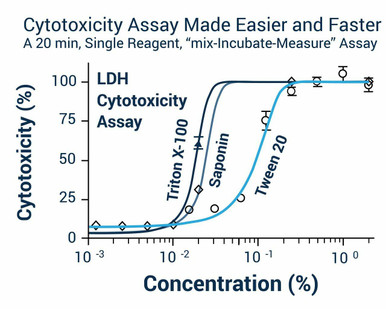
LDH Cytotoxicity Assay Kit
Detects lactate dehydrogenase released through ruptured membranes — the standard necrosis readout.
View product →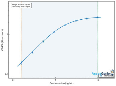
Human HMGB1 ELISA Kit
Quantifies the archetypal damage-associated molecular pattern released by necrotic cells.
View product →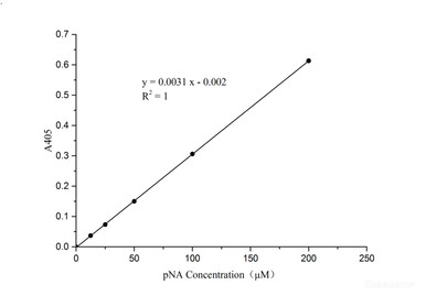
Caspase-3/7 Activity Assay Kit
Essential counterpart — low caspase activity with high LDH release points to necrosis rather than apoptosis.
View product →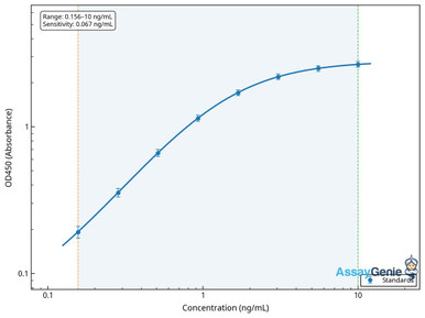
Human RIPK3 ELISA Kit
The kinase committing cells to necroptosis; its level constrains whether that pathway is even available.
View product →
Anti-MLKL Monoclonal Antibody (KO-validated)
The pore-forming executioner of necroptosis, downstream of RIPK3.
View product →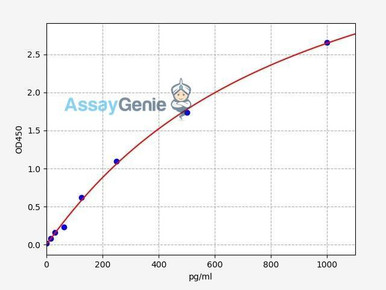
Human TNF-alpha ELISA Kit
The canonical trigger that can drive either apoptosis or necroptosis depending on cellular context.
View product →What is necrosis?
Necrosis is the death of cells or tissue within a living organism, accompanied by loss of plasma membrane integrity. That last clause carries the weight. When the membrane fails, cytoplasmic contents — enzymes, nucleotides, nuclear proteins — escape into the extracellular space, where the immune system recognises them as damage-associated molecular patterns and mounts an inflammatory response. Necrosis is therefore inherently inflammatory, and much of the tissue injury attributed to it is secondary to that inflammation rather than to the original insult.
Consequences scale with extent and location: a small focus may be cleared and replaced by scar, while extensive necrosis of myocardium, brain or kidney causes organ failure. The classical teaching contrasts necrosis, as chaotic and uncontrolled, with apoptosis, as orderly and programmed. That contrast is useful but no longer sufficient — several necrotic pathways are now known to be tightly regulated, which is covered below.
The six morphological patterns
Pathologists classify necrosis by appearance, and the pattern is informative because each associates with particular causes and tissues.
| Pattern | Appearance | Mechanism | Typical setting |
|---|---|---|---|
| Coagulative | Pale, firm; tissue architecture preserved for days | Protein denaturation outpaces enzymatic digestion | Ischaemic infarction in most solid organs, notably myocardium and kidney |
| Liquefactive | Soft, liquefied, often a cystic cavity | Enzymatic digestion dominates | Brain infarction and pyogenic bacterial abscess |
| Caseous | Granular, crumbly, cheese-like, pale yellow-white | Combined denaturation and digestion within granulomas | Tuberculosis and other granulomatous infections |
| Fat | Chalky white deposits in adipose tissue | Lipase releases free fatty acids that bind calcium (saponification) | Acute pancreatitis; traumatic injury to breast or other fat |
| Fibrinoid | Bright pink amorphous material in vessel walls | Immune complex and fibrin deposition in vessel walls | Immune-mediated vasculitis; malignant hypertension |
| Gangrenous | Blackened, shrunken (dry) or swollen and purulent (wet) | Coagulative necrosis, with superimposed bacterial liquefaction in wet gangrene | Limb ischaemia, often with diabetes or peripheral vascular disease |
Gangrene is best understood as a clinical term layered onto these patterns rather than a separate mechanism: dry gangrene is coagulative necrosis of a limb, and wet gangrene is the same with bacterial infection producing liquefaction on top. Fibrinoid necrosis is the pattern most often omitted from summaries, but it is the one that identifies immune-mediated vascular injury on biopsy.
Causes of necrosis
Anything that overwhelms a cell’s capacity to maintain ion gradients and membrane integrity can cause necrosis. The common categories:
- Ischaemia. The most frequent cause. Interrupted perfusion deprives cells of oxygen and substrate, ATP production fails, ion pumps stop, and the cell swells and ruptures. Reperfusion can paradoxically worsen injury through a burst of reactive oxygen species.
- Infection. Bacteria, viruses and fungi cause necrosis directly through toxins and enzymes, and indirectly through the immune response they provoke. Some organisms produce necrosis as their defining lesion.
- Trauma. Crush and blast injury kills cells mechanically and destroys the vasculature supplying surrounding tissue, so the necrotic zone typically exceeds the directly injured one.
- Toxins and drugs. Industrial chemicals, heavy metals, alcohol, certain therapeutics and venoms. Necrosis is often organ-selective, reflecting where a toxin is concentrated or metabolised — hence hepatic and renal tubular predominance.
- Radiation. Ionising radiation causes DNA and membrane damage directly and produces delayed necrosis through progressive vascular injury, which is why radionecrosis can appear long after exposure.
- Temperature extremes. Freezing causes ice crystal formation and vascular occlusion in frostbite; burns cause immediate coagulative necrosis.
- Immune-mediated injury. Complement activation and cytotoxic lymphocyte attack can produce necrosis, and immune complex deposition gives the fibrinoid pattern.
Regulated necrosis pathways
The most consequential change in how necrosis is understood is that a substantial part of it is programmed. These pathways produce necrotic morphology — swelling, membrane rupture, inflammation — yet run on defined molecular machinery and can be inhibited genetically or pharmacologically. For research purposes this matters enormously, because it means necrosis can be a druggable target rather than an endpoint.
Necroptosis
The best characterised regulated necrotic pathway. It is typically triggered by TNF receptor engagement under conditions where caspase-8 activity is absent or inhibited. RIPK1 and RIPK3 assemble into a complex known as the necrosome, RIPK3 phosphorylates mixed lineage kinase domain-like protein, and phosphorylated MLKL oligomerises and inserts into the plasma membrane to form pores. The cell swells and ruptures.
Two features are worth noting. Necroptosis is caspase-independent, so a pan-caspase inhibitor does not prevent it — and in fact caspase-8 inhibition promotes it, since caspase-8 normally cleaves and restrains RIPK1. And its availability depends on RIPK3 expression, which is silenced in many cultured cell lines, so a cell type that fails to necroptose may simply lack the machinery rather than being resistant.
Pyroptosis
Inflammasome-driven death, executed by inflammatory caspases — caspase-1, and caspase-4, -5 or -11 — which cleave gasdermin D. The liberated N-terminal fragment forms membrane pores, releasing mature IL-1 beta and IL-18 alongside cytoplasmic contents. It is therefore both a death mechanism and a cytokine release mechanism, and is central to antimicrobial defence and to inflammasome-driven disease.
Ferroptosis
Iron-dependent death driven by accumulation of lipid peroxides when glutathione peroxidase 4 activity is insufficient. It is morphologically necrotic but mechanistically distinct from both necroptosis and pyroptosis, requires neither caspases nor MLKL, and is inhibited by iron chelators and lipophilic antioxidants. Its role in cancer therapy resistance has made it an active target area.
Why the distinction matters experimentally
These pathways cannot be separated by membrane-integrity assays alone, because all three end in rupture and all three release lactate dehydrogenase. Discriminating them requires pathway-specific evidence: MLKL phosphorylation or RIPK3 dependence for necroptosis, gasdermin D cleavage and IL-1 beta release for pyroptosis, lipid peroxidation and rescue by an iron chelator for ferroptosis. Pharmacological rescue is the most persuasive single line of evidence — if necrostatin prevents the death, it was necroptosis.
Necrosis versus apoptosis
The two are routinely contrasted, and the contrast holds at the level of membrane integrity and inflammation even though the regulated pathways blur the older distinction between programmed and unprogrammed.
| Feature | Apoptosis | Necrosis |
|---|---|---|
| Plasma membrane | Remains intact; cell fragments into membrane-bound apoptotic bodies | Ruptures, releasing cytoplasmic contents |
| Cell volume | Shrinks | Swells before rupture |
| Inflammation | Minimal — apoptotic bodies are cleared silently by phagocytes | Prominent, driven by released damage-associated molecular patterns |
| Caspase involvement | Caspase-3 and -7 activation is central | Absent in necroptosis and ferroptosis; inflammatory caspases in pyroptosis |
| Energy requirement | ATP-dependent | Often follows ATP depletion, though regulated forms require signalling |
| Scale | Single scattered cells | Typically contiguous groups of cells or whole tissue regions |
| Laboratory readout | Caspase-3/7 activity, annexin V binding, TUNEL | Lactate dehydrogenase release, membrane-impermeant dye uptake, HMGB1 |
In practice the two overlap. The same stimulus can produce either depending on dose and cellular state — low-dose injury tends to apoptosis, high-dose to necrosis — and apoptotic cells that are not cleared undergo secondary necrosis, rupturing eventually. This is why a single-endpoint experiment is often uninterpretable, and why time course matters as much as the assay chosen.
Clinical presentations
Several named conditions are defined by necrosis in a specific tissue, and they illustrate how the same process differs by location.
| Condition | Tissue affected | Usual mechanism | Notes |
|---|---|---|---|
| Avascular necrosis (osteonecrosis) | Bone, commonly the femoral head | Interrupted blood supply | Associated with trauma, prolonged corticosteroid use, heavy alcohol intake and sickle cell disease; can progress to joint collapse |
| Acute tubular necrosis | Renal tubular epithelium | Prolonged ischaemia or nephrotoxin exposure | A leading cause of acute kidney injury; often reversible if the cause is corrected early |
| Myocardial infarction | Cardiac muscle | Coronary occlusion causing coagulative necrosis | Necrotic myocardium is replaced by non-contractile scar |
| Necrotising fasciitis | Fascia and soft tissue | Rapidly spreading bacterial infection | A surgical emergency; extent typically exceeds what is visible externally |
| Loxoscelism (recluse spider bite) | Skin and subcutaneous tissue | Sphingomyelinase D in Loxosceles venom | Necrotic ulceration is specific to recluse spiders; widow spider venom is neurotoxic and does not cause necrosis |
| Frostbite | Skin and extremities | Ice crystal formation and vascular occlusion | Damage often extends deeper than initial appearance suggests |
That last row corrects a claim worth flagging: necrotic skin lesions from spider bites are attributable to Loxosceles species, whose venom contains sphingomyelinase D. Widow spiders (Latrodectus) produce a neurotoxic envenomation with pain and muscle spasm, not tissue necrosis, and grouping the two together is a common error.
Signs, treatment and prevention
Presentation depends on site and extent, but common features of superficial necrosis include pain, discolouration progressing from pallor to dusky or black, swelling, skin that becomes tense, shiny, blistered or ulcerated, foul odour where infection is present, and loss of function in the affected part. Deep or visceral necrosis may present only with systemic signs — fever, tachycardia, organ dysfunction or raised inflammatory markers.
Management addresses the underlying cause and removes dead tissue. That typically means restoring perfusion where ischaemia is responsible, surgical debridement or amputation for extensive or infected necrosis, antimicrobials for infective causes, wound care to protect the remaining tissue, and supportive treatment for organ dysfunction. Prevention follows the causes: control of diabetes and hypertension, smoking cessation to preserve vascular health, prompt care of wounds, protective equipment in high-risk activities, and cold-weather precautions.
Suspected necrosis needs urgent medical assessment. Progression can be rapid, particularly with infection, and outcomes depend heavily on how early treatment begins. This article describes the biology for a research audience and is not clinical advice or a substitute for seeing a clinician.
How necrosis is measured
Necrosis is detected by finding evidence that membranes have failed, and confirmed by ruling out the alternative.
- Lactate dehydrogenase release. LDH is abundant and cytosolic, so its appearance in culture supernatant indicates membrane rupture. The standard first-line quantitative readout, and straightforward to normalise against a full-lysis control.
- Membrane-impermeant dyes. Propidium iodide, 7-AAD and similar dyes enter only cells with compromised membranes, allowing single-cell quantification by flow cytometry or imaging.
- Damage-associated molecular patterns. HMGB1 is the archetype — a nuclear protein released on rupture that acts as an inflammatory signal, measurable by ELISA in supernatant, serum or plasma.
- Caspase-3/7 activity. Included to exclude apoptosis rather than to detect necrosis. High LDH with low caspase activity is the signature of primary necrosis; both high suggests apoptosis proceeding to secondary necrosis.
- Pathway-specific markers. Phosphorylated MLKL for necroptosis, cleaved gasdermin D and IL-1 beta for pyroptosis, lipid peroxidation for ferroptosis.
- Histology. On tissue, morphology remains definitive — the six patterns above are recognised on routine stains, and TUNEL distinguishes apoptotic nuclei.
Pair at least one membrane-integrity readout with one caspase readout, run a full-lysis control for normalisation, and take more than one timepoint. A single endpoint cannot distinguish primary necrosis from apoptosis that has progressed to rupture.
Tumour necrosis factor and necrosis
TNF-alpha was named for its ability to induce tumour necrosis, and the relationship remains instructive. It is produced mainly by macrophages and T cells, and on binding TNF receptor 1 it assembles a signalling complex whose output depends on cellular context rather than on the ligand.
Three outcomes are possible from the same receptor. Most commonly TNF drives NF-kappa-B activation and survival with inflammatory gene expression. Where NF-kappa-B signalling is blocked, caspase-8 is activated and the cell undergoes apoptosis. Where caspase-8 is additionally absent or inhibited, RIPK3 and MLKL are engaged instead and the cell undergoes necroptosis. TNF is therefore the standard experimental trigger for necroptosis, applied together with a SMAC mimetic and a caspase inhibitor, and it explains why the same cytokine appears as protective in one system and lethal in another.
Choosing a cell death assay
LDH cytotoxicity and caspase activity assays, HMGB1 and TNF-alpha ELISA kits, and antibodies against the necroptosis machinery — for distinguishing necrosis from apoptosis rather than just detecting death.
Browse cell death assays →Frequently asked questions
How does necrosis differ from apoptosis?
Apoptosis keeps the plasma membrane intact, packaging the cell into bodies cleared quietly by phagocytes, and depends on caspase-3 and -7. Necrosis involves membrane rupture and release of cytoplasmic contents, which provokes inflammation. Apoptosis affects scattered single cells; necrosis usually affects contiguous groups.
Is necrosis always uncontrolled?
No, and this is the main way older descriptions are out of date. Necroptosis, pyroptosis and ferroptosis are genetically defined pathways with necrotic morphology, each with its own machinery, and each can be blocked pharmacologically.
What is tumour necrosis factor?
A cytokine produced chiefly by macrophages and T cells. Through TNF receptor 1 it can drive survival, apoptosis or necroptosis depending on whether NF-kappa-B signalling and caspase-8 are functional — which is why the same cytokine looks protective in some systems and lethal in others.
Which assay should I use to detect necrosis?
Start with lactate dehydrogenase release or a membrane-impermeant dye, and pair it with a caspase-3/7 assay. High membrane permeability with low caspase activity indicates necrosis; both elevated suggests apoptosis progressing to secondary rupture.
Why does caspase inhibition sometimes increase cell death?
Because caspase-8 normally restrains RIPK1. Inhibiting caspases removes that brake, so cells exposed to TNF are diverted from apoptosis into necroptosis instead of surviving. This is the basis of the standard experimental protocol for inducing necroptosis.
Do all spider bites cause necrosis?
No. Necrotic skin lesions are associated with recluse spiders (Loxosceles), whose venom contains sphingomyelinase D. Widow spider venom is neurotoxic, causing pain and muscle spasm rather than tissue necrosis.
What are the six morphological types of necrosis?
Coagulative, liquefactive, caseous, fat, fibrinoid and gangrenous. Fibrinoid is the one most often left out of summaries, and it is the pattern that identifies immune-mediated vascular injury.
Lauryn McLoughlin completed her undergraduate degree in Neuroscience before completing her masters in Biotechnology at University College Dublin.
Recent Posts
-
Suspected Plague Death at a Siberian Anti-Plague Institute: What Researchers Should Know
Infectious Disease · News AnalysisSuspected Plague Death at a Siberian Anti-Plague Institute: What …6th Oct 2026 -
LPS ELISA Troubleshooting: The Importance of Sample Nature
Quick answer A researcher measuring circulating lipopolysaccharide (LPS) in human serum …25th Sep 2026 -

Sekundärantikörper wählen: Die Regel, die im Labor wirklich zählt
Sekundärantikörper wählen: Die Regel, die im Labor wirklich zählt Wählen Sie einen Sekundärantikörp …24th Sep 2026