
Insulin Signaling and RTK: An Overview
Insulin Signalling & Receptor Tyrosine Kinases
Insulin controls metabolism and growth through the insulin receptor — a receptor tyrosine kinase that branches into the metabolic PI3K–AKT arm and the mitogenic RAS–MAPK/ERK arm. This guide covers the receptors, cascades, negative regulators and disease links.
Browse Signalling Antibodies →Quick answer
Insulin signalling begins when insulin binds the insulin receptor — a receptor tyrosine kinase (RTK) — triggering autophosphorylation and recruitment of IRS adaptor proteins. Signalling then branches into two arms: the PI3K–AKT pathway, which drives glucose uptake via GLUT4 and metabolic control, and the RAS–MAPK/ERK pathway, which governs growth and gene expression. Disrupted insulin signalling underlies type 2 diabetes, atherosclerosis and cancer.
Insulin-signalling antibodies
Probe the insulin pathway from receptor to output with validated phospho-specific and total antibodies against its core nodes.
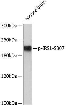
Phospho-IRS1 (S307) Antibody
Detects IRS-1 serine-307 phosphorylation, a key marker of insulin resistance.
View antibody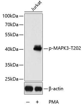
Phospho-ERK1 (T202) Antibody
Detects activated ERK1, the mitogenic output of the RAS–MAPK arm.
View antibody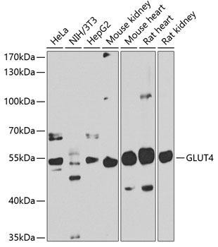
GLUT4 Antibody
Detects the insulin-responsive glucose transporter GLUT4, the metabolic endpoint.
View antibodyKey Takeaways:
- Insulin signaling involves receptor activation, signal transduction, and gene expression.
- Insulin receptors, a type of Receptor Tyrosine Kinase (RTK), regulate metabolism and cell responses.
- The Ras/MAP kinase and PI3K pathways are crucial in insulin signaling.
- Insulin signaling impacts atherosclerosis and cancer, with RTK inhibitors showing therapeutic potential.
Insulin Signaling
The insulin signalling pathway is a series of steps that occur when insulin binds to its receptor on the surface of a cell. This results in the activation of various proteins, which then initiate a range of metabolic processes.
The insulin signalling pathway can be divided into three main stages:
- Receptor activation
- Downstream signal transduction
- Target gene expression
The insulin receptor is a heterotetrameric complex of two α-subunits and two β-subunits that are disulfide linked. The insulin molecule binds to the extracellular α-subunits, which activate the intracellular tyrosine kinase domain of the β-subunit.
Insulin is a peptide hormone produced by the beta-cells of the pancreas that regulates human metabolism. Insulin binds to its receptor, causing a triphosphorylation of the activation loop. The kinase then phosphorylates tyrosine amino acids outside the kinase domain of the receptor, creating binding sites for signaling protein partners with SH2 (src-homology 2) domains or PTB (phosphotyrosine-binding) domains. The insulin receptors interact with the phosphorylated juxtamembrane domain residue of large docking proteins called IRS (Insulin receptor substrate) and the adapter Sh2 (Src homology 2 domain containing). The protein binds to and activates other proteins, such as SHC (src homology domain containing) and Grb-14 (growth factor receptor bound), which are part of the receptor complex. Following this, two pathways are activated: the Ras/MAP kinase pathway and the PI-kinase pathway.
Receptor Tyrosine Kinase (RTKs)
The insulin receptor is a tyrosine kinase receptor (RTK) that is activated by the insulin hormone. The insulin RTK pathway regulates cellular responses to insulin, such as glucose uptake, membrane potential, protein tyrosine phosphorylation and glycolysis. The first step in the pathway is activated by binding of insulin to the insulin receptor. The second step is initiated with tyrosine kinase activity, which phosphorylates serine and threonine residues on proteins and initiates a cascade of intracellular events. This cascade then leads to gene expression, protein synthesis and cell growth.
Receptor Tyrosine Kinases (RTK) Pathway
The structure of RTKs consist of an extracellular ligand-binding domain, a single transmembrane helix, and an intracellular region that includes a juxtamembrane regulatory area (TKD), as well as a tyrosine kinase domain (TKD) and a carboxyl terminal tail. RTK plays an important role in insulin signalling, which is the process by which insulin regulates the metabolism of glucose and other nutrients
There are four main types of RTKs: insulin receptor, IGF-I receptor (insulin-like growth factor I receptor), PDGF receptor (platelet derived growth factor receptor) and EGFR receptor (epidermal growth factor receptor). These receptors play an important role in regulating various aspects of cellular function, including proliferation, differentiation and survival. The insulin receptor is the most well studied RTK, and has been shown to play a role in cancer progression.
Related Resources
The RAS/MAPK-ERK Signaling Pathway
The Ras/MAP kinase pathway is activated by the insulin signaling pathway. The Ras/MAP kinase pathway is responsible for transmitting the signal from the receptor to the nucleus of the cell and the activation of a protein called Raf through the activity of Grb-14. Raf then activates two proteins called MEK and ERK, enter the nucleus and phosphorylate various transcription factors, which activate gene expression for genes that regulate metabolism. MEK, which in turn activates a protein called MAP kinase. The MAP kinase (MAPK) pathway plays an important role in regulating cell growth and survival.
Once the insulin receptor is active, the adaptor proteins Shc and Grb2 recruit the guanine-nucleotide exchange factor SOS to the membrane. SOS converts inactive RAS-GDP into active RAS-GTP, which nucleates a kinase cascade: RAS activates RAF, RAF phosphorylates MEK1/2, and MEK activates ERK1/2. Activated ERK then translocates to the nucleus, where it phosphorylates transcription factors such as ELK1 to drive proliferation, differentiation and survival programmes — the mitogenic counterpart to the metabolic PI3K–AKT branch.
The PI3K Signaling Pathway
The PI-kinase pathway is activated by the insulin signaling pathway. The PI-kinase pathway is responsible for the activation of a protein called PI-kinase and for relaying the signal from the receptor to the interior of the cell. This protein then phosphorylates other proteins, including PKB (protein kinase B) and PDK-I (phosphoinositide dependent kinase I),which regulates glucose metabolism. Phosphorylation of these proteins results in their activation, which leads to the regulation of various metabolism.
Once activated, PKB and PDK-I promote the fusion of GLUT-IV (glucose transporter type IV) with the cell membrane. GLUT-IV transports glucose into the cell, and GS (glucagon receptor), which regulates blood sugar levels. This allows glucose to enter the cell and be used for energy.
Negative Regulators of Insulin Signalling
There are a number of proteins that act as negative regulators of insulin signalling. These proteins help to prevent the receptor from being activated too strongly and causing excessive cell proliferation. Some of the most well studied negative regulators of insulin signalling include PTEN (phosphatase and tensin homolog), FOXO (forkhead box O) and SREBP-lc (sterol regulatory element binding protein-lc).
PTEN is a lipid and protein tyrosine phosphatase that dephosphorylates the receptor, leading to its inactivation. PTEN is a tumor suppressor gene, and mutations in this gene are associated with various types of cancer.
FOXO is a transcription factor that regulates the expression of genes involved in metabolism and cell growth. FOXO has been shown to inhibit insulin signalling by blocking the activation of PKB.
SREBP-lc is a transcription factor that regulates the synthesis of cholesterol and other lipids. SREBP-lc has been shown to promote insulin resistance by inhibiting PKB activity.
Insulin Related Products
The Role of Insulin Signalling in Atherosclerosis
Atherosclerosis is a complex pathological process that involves a variety of cells, including vascular, immune, and metabolic cells. Insulin signaling via the insulin receptors plays an essential role in vascular cells and vessel dilation and relaxation.
Increasing evidence suggests that hyperinsulinemia is linked to the development of atherosclerosis in diabetes. This is supported by human studies showing that insulin infusion in healthy persons causes vasodilation and improves blood flow to the peripheral tissues. These effects are dampened in those with insulin resistance and type 2 diabetes.
The receptor tyrosine kinase inhibitors (RTKIs) sunitinib, lapatinib and gefitinib have all been shown to reduce lesion size and inhibit foam cell formation in mice models of atherosclerosis.
The Role of Insulin Signalling in Cancer
There is growing evidence that receptor tyrosine kinases play a role in the development of cancer. Evidence suggests that Insulin receptors are over-expressed in cancer cells, especially those of the breast cancer. Increased insulin receptor expression is associated with shorter survival in breast cancers. Insulin receptors are frequently over-expressed and highly phosphorylated in breast tumors generated by diabetic mice.
The receptor tyrosine kinase inhibitors (RTKIs) sunitinib, lapatinib and gefitinib have all been shown to reduce tumor size and inhibit cell proliferation in various types of cancer cells.
Seán Mac Fhearraigh PhD is a co-founder of Assay Genie. Seán carried out his undergraduate degree in Genetics at Trinity College Dublin, followed by a PhD at University College Dublin. He carried out a post-doc at the Department of Genetics, University of Cambridge. Seán is now Chief Technical Officer at Assay Genie.
Frequently asked questions
What is the difference between the PI3K and MAPK branches of insulin signalling?
The PI3K–AKT branch is largely metabolic — promoting GLUT4-mediated glucose uptake, glycogen synthesis and protein synthesis. The RAS–MAPK/ERK branch is mitogenic, regulating growth, proliferation and gene expression. Both originate at the activated insulin receptor and IRS proteins.
What is insulin resistance?
Insulin resistance is a reduced cellular response to insulin, often driven by chronic serine phosphorylation of IRS-1 (e.g. at Ser307) and impaired PI3K–AKT signalling. It blunts GLUT4 translocation and glucose uptake and is central to type 2 diabetes and metabolic syndrome.
Why is the insulin receptor a receptor tyrosine kinase?
The insulin receptor is a transmembrane enzyme whose intracellular domain phosphorylates tyrosine residues. Insulin binding triggers autophosphorylation of the β-subunits, creating docking sites for IRS and Shc adaptors that propagate the signal.
How is insulin signalling studied in the lab?
Researchers use phospho-specific antibodies against key nodes (phospho-insulin receptor, phospho-IRS-1, phospho-AKT, phospho-ERK) in Western blot or ELISA, alongside glucose-uptake and GLUT4-translocation assays, to quantify pathway activity.
Related articles
Researching cell signalling?
From phospho-specific antibodies to glucose and metabolism assay kits, Assay Genie supports signal-transduction research with publication-ready reagents and expert technical support.
Browse Signalling Antibodies →Recent Posts
-
Suspected Plague Death at a Siberian Anti-Plague Institute: What Researchers Should Know
Infectious Disease · News AnalysisSuspected Plague Death at a Siberian Anti-Plague Institute: What …6th Oct 2026 -
LPS ELISA Troubleshooting: The Importance of Sample Nature
Quick answer A researcher measuring circulating lipopolysaccharide (LPS) in human serum …25th Sep 2026 -

Sekundärantikörper wählen: Die Regel, die im Labor wirklich zählt
Sekundärantikörper wählen: Die Regel, die im Labor wirklich zählt Wählen Sie einen Sekundärantikörp …24th Sep 2026