
A Guide To Tau Proteins & Tauopathies
A Guide to Tau Proteins & Tauopathies
Tau keeps the neuronal cytoskeleton standing up. When its phosphorylation goes wrong, the same protein becomes the aggregate that defines Alzheimer’s disease, frontotemporal dementia and progressive supranuclear palsy. This guide covers tau’s structure and isoforms, the disorders it drives, and how researchers measure it.
Browse tau assays →Key takeaways
- Tau, encoded by MAPT on chromosome 17, binds and stabilises axonal microtubules through its repeat domains.
- Alternative splicing yields six CNS isoforms, split into three-repeat (3R) and four-repeat (4R) classes.
- Physiological phosphorylation tunes microtubule binding; hyperphosphorylation detaches tau and drives it into paired helical filaments and neurofibrillary tangles.
- Tauopathies are grouped by which repeat class dominates the aggregate, which is why they differ in anatomy and symptoms.
- Tau is also expressed outside the nervous system, at much lower abundance, where it contributes to cell division and differentiation.
- Total tau, phospho-tau, amyloid-beta, NfL and GFAP are typically measured together rather than in isolation.
Contents
- What are tau proteins?
- The structure of tau
- Where tau is found
- What are tauopathies?
- Tau in Alzheimer’s disease
- Frontotemporal dementia
- Progressive supranuclear palsy
- Parkinson’s disease
- Comparing the major tauopathies
- Tau and Down syndrome
- Tau kinases
- Mutation versus phosphorylation
- How tau is measured
- Choosing a tau assay
- Frequently asked questions
Tau & neurodegeneration assay kits
A tau experiment rarely stands alone. The panel below pairs total and phosphorylated tau with the amyloid, axonal-injury and astroglial markers most often profiled alongside them.
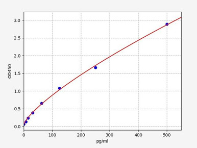
Human MAPT / Microtubule-Associated Protein Tau ELISA Kit
Quantifies total tau across CSF, serum, plasma and tissue lysates — the baseline measure of tau burden.
View kit →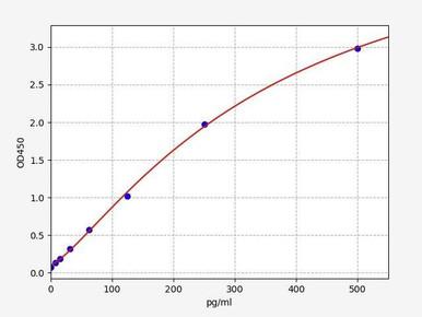
Human Phospho-Tau (Thr181) ELISA Kit
Targets the Thr181 phospho-epitope that underpins current blood-based Alzheimer’s staging work.
View kit →
Human Beta-Amyloid (1-42) ELISA Kit
Resolves the amyloid arm of Alzheimer’s pathology alongside tau readouts.
View kit →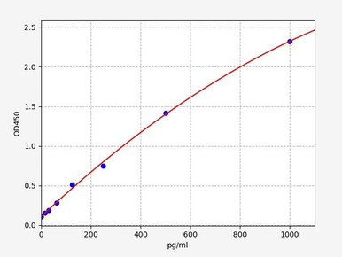
Human NEFL / Neurofilament Light ELISA Kit
Neurofilament light reports axonal damage across tauopathies and other neurodegenerative disease.
View kit →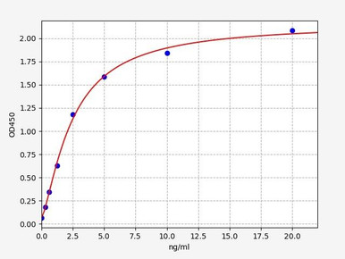
Human GFAP ELISA Kit
Astrocytic reactivity marker, routinely co-profiled with tau and neurofilament light.
View kit →
Human GSK-3β / Glycogen Synthase Kinase-3 Beta ELISA Kit
Measures the principal tau kinase implicated in pathological hyperphosphorylation.
View kit →What are tau proteins?
Tau proteins are a family of microtubule-associated proteins that maintain the internal scaffolding of neurons. The best characterised member, microtubule-associated protein tau, binds along microtubules and promotes their assembly while resisting their disassembly. Microtubules matter because they are the tracks along which cargo is trafficked down the axon, and the framework that lets a neuron hold an elaborate shape over decades.
In a healthy neuron tau is concentrated in the axon, dynamically bound to microtubules, and its phosphorylation state is tightly regulated. That regulation is the point of failure. In Alzheimer’s disease and related disorders, tau becomes abnormally modified, releases the microtubule surface, and self-assembles into paired helical filaments that mature into neurofibrillary tangles. The cytoskeleton it once stabilised then destabilises, transport fails, and the neuron degenerates.
The structure of tau
Tau is encoded by the MAPT gene on the long arm of chromosome 17. The six isoforms expressed in adult human CNS span roughly 45–65 kDa, and tau accounts for only a small fraction of total brain protein despite its outsized pathological importance. Structurally, tau is an intrinsically disordered protein organised into functional regions:
- The N-terminal projection domain, which does not bind microtubules directly but projects away from the microtubule surface and sets spacing between adjacent filaments. Alternative splicing of exons 2 and 3 inserts zero, one or two N-terminal sequences, giving the 0N, 1N and 2N variants.
- The proline-rich mid-region, densely populated with serine and threonine residues and the main substrate for tau kinases.
- The microtubule-binding repeat region, in the C-terminal half, comprising either three or four imperfect repeats depending on whether exon 10 is included.
- The C-terminal tail, which carries further phosphorylation sites and contributes to filament assembly.
Because exon 10 splicing determines repeat number, the six isoforms sort into two classes: three-repeat (3R) and four-repeat (4R) tau. Four-repeat tau binds microtubules more avidly than three-repeat tau. Which class dominates an aggregate is one of the most useful ways to classify a tauopathy, and adult brain normally holds the two in approximate balance.
Where tau is found
Tau is overwhelmingly a nervous-system protein, most abundant in the axons of CNS neurons, but it is not exclusive to them. Lower levels appear in peripheral tissues including kidney, heart, lung and skeletal muscle, where tau participates in cell division and differentiation and so contributes to tissue development, maintenance and repair. The abundance gap is large enough that peripheral tau rarely dominates a measurement of circulating tau.
MAPT variation has also been linked to disease outside the classical tauopathies, including some cancers, where altered tau function has been proposed to affect microtubule dynamics in dividing cells and the response to microtubule-targeting drugs.
What are tauopathies?
Tauopathies are neurodegenerative disorders defined by the abnormal accumulation of aggregated tau in the brain. They present with some combination of cognitive decline, behavioural and personality change, and motor impairment, with the mix determined by which cells and regions accumulate tau first. Alzheimer’s disease is by far the most common, but the family also includes frontotemporal lobar degeneration with tau pathology, progressive supranuclear palsy, corticobasal degeneration, Pick’s disease and chronic traumatic encephalopathy.
A practical distinction separates primary tauopathies, where tau aggregation is the leading pathology, from secondary tauopathies such as Alzheimer’s disease, where tau pathology accompanies another driver — in this case amyloid-beta. The distinction matters for therapeutic strategy: in a primary tauopathy, tau is the target.
Tau in Alzheimer’s disease
Alzheimer’s disease is a progressive neurodegenerative disorder that principally erodes memory and cognition, and it is the most common cause of dementia worldwide. Two pathologies define it: extracellular amyloid-beta plaques and intracellular neurofibrillary tangles built from hyperphosphorylated tau containing both 3R and 4R isoforms.
Tau pathology in Alzheimer’s follows a strikingly stereotyped anatomical progression. It begins in the entorhinal cortex and transentorhinal region, spreads to the hippocampus, and then extends across association cortices — the sequence formalised as Braak staging. That spatial pattern tracks cognitive decline far more closely than plaque distribution does, which is a large part of why tau attracted such intense interest as both biomarker and drug target. Clinically the result is progressive memory loss, disorientation, and difficulty with language and problem-solving.
Frontotemporal dementia
Frontotemporal dementia describes a group of disorders in which the frontal and temporal lobes degenerate, often with onset earlier than in Alzheimer’s disease. Roughly half of frontotemporal lobar degeneration cases carry tau pathology, with the remainder driven mostly by TDP-43 or FUS. Where tau is the culprit, the aggregate composition varies by subtype: Pick’s disease is a 3R tauopathy with characteristic Pick bodies, while other subtypes are predominantly 4R.
Because the damage lands on circuits governing behaviour, personality and language, the presentation differs sharply from the amnestic picture of Alzheimer’s. Patients may show disinhibition or apathy, emotional blunting, loss of empathy, progressive aphasia, and executive dysfunction, while memory can remain comparatively preserved early on. Causative mutations in MAPT itself were first identified in inherited frontotemporal dementia with parkinsonism, which established that tau dysfunction alone is sufficient to cause neurodegeneration.
Progressive supranuclear palsy
Progressive supranuclear palsy is a rare primary 4R tauopathy in which tau accumulates in both neurons and glia across the basal ganglia, brainstem, cerebellar nuclei and parts of the cortex. Its tufted astrocytes are a diagnostic hallmark on neuropathology.
The clinical signature is motor: early postural instability with backward falls, axial rigidity, and a vertical supranuclear gaze palsy that gives the condition its name. Cognitive change follows a frontal-executive pattern rather than an amnestic one, with impaired attention, reduced verbal fluency and slowed processing. Because it is a primary tauopathy with a relatively homogeneous pathology, progressive supranuclear palsy has become a favoured setting for testing tau-directed therapeutics.
Parkinson’s disease
Parkinson’s disease is defined by progressive loss of dopaminergic neurons in the substantia nigra and by alpha-synuclein aggregation into Lewy bodies, so it is a synucleinopathy rather than a tauopathy. Tau still appears in the story, however. Tau co-deposits with alpha-synuclein in a substantial minority of cases, MAPT haplotype variation is an established genetic risk factor for Parkinson’s, and the two proteins appear to promote one another’s aggregation experimentally.
Cases with mixed tau and synuclein pathology tend toward more pronounced cognitive impairment than classical Parkinson’s. The motor syndrome itself — resting tremor, bradykinesia, rigidity and postural instability — is accompanied by non-motor features including cognitive decline, mood disturbance, disrupted sleep and autonomic dysfunction. Untangling how tau and alpha-synuclein interact remains an active research question.
Comparing the major tauopathies
The disorders above overlap enough to be confused clinically and differ enough that the distinction changes management and research design. The table below sets the four side by side.
| Feature | Alzheimer’s disease | Frontotemporal dementia (tau) | Progressive supranuclear palsy | Parkinson’s disease |
|---|---|---|---|---|
| Defining aggregate | Amyloid-beta plaques plus tau tangles | Tau inclusions (or TDP-43 in non-tau subtypes) | Neuronal and glial tau, tufted astrocytes | Alpha-synuclein Lewy bodies |
| Tau repeat class | Mixed 3R and 4R | 3R in Pick’s disease, 4R in several other subtypes | 4R | Not primary; tau may co-deposit |
| First regions affected | Entorhinal cortex and hippocampus | Frontal and temporal lobes | Basal ganglia, brainstem, cerebellar nuclei | Substantia nigra |
| Leading features | Memory loss, disorientation, language and problem-solving difficulty | Behavioural change, disinhibition or apathy, aphasia | Falls, axial rigidity, vertical gaze palsy | Resting tremor, bradykinesia, rigidity |
| Typical decline | Gradual over years | Often faster than Alzheimer’s | Rapid, with early loss of mobility | Variable; faster cognitive decline when tau co-occurs |
| Is tau the driver? | Secondary to amyloid-beta | Yes, in tau-positive subtypes | Yes | No |
Two comparisons come up repeatedly. Against Parkinson’s disease, the separation is aggregate and anatomy: Alzheimer’s is amyloid and tau in medial temporal cortex producing an amnestic syndrome, Parkinson’s is alpha-synuclein in the substantia nigra producing a movement disorder. Against frontotemporal dementia, both are tau-driven cortical diseases, but frontotemporal dementia strikes frontal and temporal circuits first, so behaviour and language fail before memory does, and onset is typically earlier. Neither Alzheimer’s disease nor frontotemporal dementia is currently curable, though symptomatic treatment and, for Alzheimer’s, anti-amyloid immunotherapy can modify the course.
Tau and Down syndrome
Down syndrome carries an extra copy of chromosome 21, which is also where the amyloid precursor protein gene sits. Lifelong overexpression of amyloid precursor protein produces amyloid-beta accumulation from early adulthood, and tau pathology follows, giving a very high lifetime incidence of Alzheimer’s-type dementia — the great majority of people with Down syndrome show Alzheimer’s neuropathology by their sixties.
This makes Down syndrome one of the clearest natural demonstrations that amyloid-beta accumulation can precede and precipitate tau pathology, and it has made the population important to studies of biomarker sequence and of early intervention. Understanding the interplay between amyloid, tau and cognition here informs therapeutic strategy well beyond Down syndrome itself.
Tau kinases
A tau kinase is an enzyme that transfers phosphate groups onto specific serine, threonine or tyrosine residues on tau. Tau carries an unusually large number of potential phosphorylation sites — more than eighty across the full-length protein — and their occupancy determines how tightly tau holds microtubules. Modest, reversible phosphorylation is how a neuron loosens tau to remodel its cytoskeleton.
Dysregulation converts that control system into a disease mechanism. When kinase activity outruns the opposing phosphatases, principally protein phosphatase 2A, tau becomes hyperphosphorylated, detaches, and aggregates. The kinases most implicated are glycogen synthase kinase-3 beta, cyclin-dependent kinase 5, the mitogen-activated protein kinases, protein kinase A, and the microtubule-affinity-regulating kinases. Glycogen synthase kinase-3 beta has drawn the most attention because it phosphorylates many of the epitopes found in Alzheimer’s tangles, which is why kinase inhibition has been pursued repeatedly as a therapeutic strategy.
Mutation versus phosphorylation
These two routes to tau pathology are easy to conflate but are mechanistically distinct. A tau mutation is a heritable change in the MAPT sequence. Mutations act either by altering the protein directly, weakening microtubule binding and raising aggregation propensity, or by shifting exon 10 splicing so that the 3R to 4R ratio is disturbed. Such mutations cause inherited frontotemporal dementia with parkinsonism and related syndromes, and they are the reason tau is considered a primary cause of neurodegeneration rather than a bystander.
Tau phosphorylation, by contrast, is a reversible post-translational modification present in every healthy neuron. It is not inherently harmful — it is the normal mechanism regulating tau’s affinity for microtubules. The pathological state is hyperphosphorylation: abnormally high stoichiometry at abnormal sites, which detaches tau from microtubules, raises the free cytosolic pool, and favours assembly into paired helical filaments. Most sporadic tauopathy involves hyperphosphorylation without any MAPT mutation at all.
How tau is measured
Measuring tau serves two purposes: diagnosing and staging disease in people, and quantifying tau in experimental systems. The available methods differ in what they can see and when they can see it.
Tau PET imaging
Positron emission tomography with a tau-selective radiotracer visualises aggregated tau in the living brain and can be repeated longitudinally. Tracer binding maps closely onto Braak staging, which makes tau PET valuable for tracking spread over time. It is expensive, requires specialist infrastructure, and detects aggregates rather than soluble tau species.
Cerebrospinal fluid and blood biomarkers
Immunoassays quantify soluble tau in biofluids. Total tau in cerebrospinal fluid reflects general neuronal injury, while phosphorylated tau is far more specific to Alzheimer’s pathology. The shift over the last decade has been from cerebrospinal fluid to plasma: phospho-tau measured at Thr181, Thr217 or Thr231 is now detectable in blood at concentrations that discriminate Alzheimer’s disease from other dementias, which removes the need for a lumbar puncture. Tau is rarely measured alone — amyloid-beta 42, neurofilament light as a marker of axonal damage, and glial fibrillary acidic protein as a marker of astrocytic reactivity are typically profiled in the same sample to separate tau-specific change from generalised neurodegeneration.
Tissue and cell-based methods
Post-mortem neuropathology remains the reference standard. Immunohistochemistry and immunofluorescence localise tau and its aggregates to particular cells and layers, allowing Braak staging and identification of features such as tufted astrocytes or Pick bodies. Western blotting resolves tau isoforms and phospho-species by molecular weight, and is how the 3R versus 4R composition of an aggregate is established. Sandwich ELISA remains the workhorse for quantifying total or phosphorylated tau in lysates, conditioned media and biofluids, and pairs naturally with kinase measurements when the question concerns phosphorylation state.
What this means clinically
There is no treatment that reverses a tau mutation, and no disease-modifying therapy directed at tau has yet been approved. Management focuses on symptomatic treatment, monitoring, and — for Alzheimer’s disease specifically — anti-amyloid immunotherapy, alongside cardiovascular risk control, exercise, sleep and cognitive engagement, which are associated with slower decline. Anyone with a known pathogenic MAPT variant or a strong family history should be followed by a specialist, and this article is not a substitute for that advice.
Choosing a tau assay
Total tau, phospho-tau, amyloid-beta, neurofilament light and GFAP kits validated for human CSF, serum, plasma and lysates — with matched kinase assays for phosphorylation studies.
Browse tau & neurodegeneration kits →Frequently asked questions
Is Alzheimer’s disease a tauopathy?
Yes, though a secondary one. Tau tangles are one of its two defining pathologies, but amyloid-beta accumulation is generally considered the upstream driver. Primary tauopathies such as progressive supranuclear palsy have tau aggregation as their leading pathology.
What is the difference between 3R and 4R tau?
Whether exon 10 of MAPT is included determines whether tau carries three or four microtubule-binding repeats. Four-repeat tau binds microtubules more tightly. Which class dominates an aggregate helps classify the tauopathy: progressive supranuclear palsy is 4R, Pick’s disease is 3R, and Alzheimer’s disease contains both.
Why is phospho-tau a better Alzheimer’s biomarker than total tau?
Total tau rises with neuronal injury of almost any cause, so it is sensitive but not specific. Phosphorylated tau, particularly at Thr181, Thr217 and Thr231, tracks Alzheimer’s pathology specifically and can now be measured in plasma rather than cerebrospinal fluid.
Can tau be measured in blood?
Yes. Plasma phospho-tau assays have improved to the point that they discriminate Alzheimer’s disease from other dementias without a lumbar puncture, and they are usually run alongside neurofilament light and GFAP to separate tau-specific change from general neurodegeneration.
Does hyperphosphorylated tau cause neurodegeneration, or result from it?
Both directions have evidence. Hyperphosphorylation detaches tau from microtubules and promotes aggregation, which impairs axonal transport — a causal route. But cellular stress and failing phosphatase activity also increase phosphorylation, so it amplifies as disease progresses.
Which sample types work with tau ELISA kits?
Sandwich ELISA kits for tau are typically validated for serum, plasma, cerebrospinal fluid, cell culture supernatant and tissue lysates. Always check the validated matrix and dynamic range on the individual kit datasheet before designing an experiment.
Lauryn McLoughlin completed her undergraduate degree in Neuroscience before completing her masters in Biotechnology at University College Dublin.
Recent Posts
-
Suspected Plague Death at a Siberian Anti-Plague Institute: What Researchers Should Know
Infectious Disease · News AnalysisSuspected Plague Death at a Siberian Anti-Plague Institute: What …6th Oct 2026 -
LPS ELISA Troubleshooting: The Importance of Sample Nature
Quick answer A researcher measuring circulating lipopolysaccharide (LPS) in human serum …25th Sep 2026 -

Sekundärantikörper wählen: Die Regel, die im Labor wirklich zählt
Sekundärantikörper wählen: Die Regel, die im Labor wirklich zählt Wählen Sie einen Sekundärantikörp …24th Sep 2026