The BRSK1 Antibody (CAB17226) is a high-quality antibody developed for reliable detection and analysis of target proteins. Enables magnesium ion binding activity; protein serine/threonine kinase activity; and tau-protein kinase activity. Involved in mitotic G2 DNA damage checkpoint signaling and protein phosphorylation. Acts upstream of or within G2/M transition of mitotic cell cycle; peptidyl-serine phosphorylation; and response to UV. Located in cell junction; cytoplasm; and nucleoplasm. RRID AB_2768626 Gene ID 84446 Swiss Prot Synonym SAD-B; hSAD1; BRSK1
This antibody is validated for use in WB, IHC-P, ELISA applications and has demonstrated reactivity against Human, Mouse, Rat samples.
Product Name:
BRSK1 Antibody
SKU:
CAB17226
Size:
100μL, 20μL
Reactivity:
Human, Mouse, Rat
Clone Number:
-
Conjugate:
Unconjugated
Immunogen:
Recombinant protein (or fragment).This information is considered to be commercially sensitive.
Tested Applications:
WBIHC-PELISA
Recommended Dilution:
WB
1:500 - 1:1000
IHC-P
1:50 - 1:200
ELISA
Recommended starting concentration is 1 μg/mL. Please optimize the concentration based on your specific assay requirements.
Synonyms:
SAD-B, hSAD1, BRSK1
Positive Sample:
Raji
Cellular Localization:
Cell Junction, Cytoplasm, Nucleus, Centrosome, Cytoskeleton, Microtubule Organizing Center, Synapse.
Calculated MW:
85kDa
Observed MW:
90kDa
Enables magnesium ion binding activity; protein serine/threonine kinase activity; and tau-protein kinase activity. Involved in mitotic G2 DNA damage checkpoint signaling and protein phosphorylation. Acts upstream of or within G2/M transition of mitotic cell cycle; peptidyl-serine phosphorylation; and response to UV. Located in cell junction; cytoplasm; and nucleoplasm. RRID AB_2768626 Gene ID 84446 Swiss Prot Synonym SAD-B; hSAD1; BRSK1
Purification Method:
Affinity purification
Gene ID:
84446
RRID:
AB_2768626
Buffer Information:
Store at -20℃. Avoid freeze / thaw cycles. Buffer: PBS with 0.01% thimerosal,50% glycerol,pH7.3.
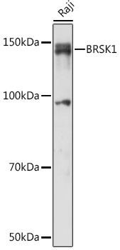
Western blot analysis of lysates from Raji cells, using BRSK1 Rabbit pAb (CAB17226) at 1:1000 dilution. Secondary antibody: HRP-conjugated Goat anti-Rabbit IgG (H+L) (AS014) at 1:10000 dilution. Lysates/proteins: 25μg per lane. Blocking buffer: 3% nonfat dry milk in TBST. Detection: ECL Basic Kit (AbGn00020). Exposure time: 60s.
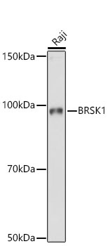
Western blot analysis of lysates from Raji cells using BRSK1 Rabbit pAb (CAB17226) at 1:1000 dilution. Secondary antibody: HRP-conjugated Goat anti-Rabbit IgG (H+L) (AS014) at 1:10000 dilution. Lysates/proteins: 25 μg per lane. Blocking buffer: 3% nonfat dry milk in TBST. Detection: ECL Basic Kit (AbGn00020). Exposure time:10s.
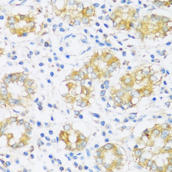
Immunohistochemistry analysis of paraffin-embedded Human stomach using BRSK1 Rabbit pAb (CAB17226) at dilution of 1:100 (40x lens). Microwave antigen retrieval performed with 0.01M PBS Buffer (pH 7.2) prior to IHC staining.
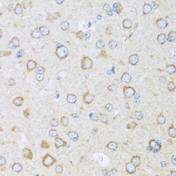
Immunohistochemistry analysis of paraffin-embedded Mouse brain using BRSK1 Rabbit pAb (CAB17226) at dilution of 1:100 (40x lens). Microwave antigen retrieval performed with 0.01M PBS Buffer (pH 7.2) prior to IHC staining.