The BYSL Antibody (CAB11993) is a high-quality antibody developed for reliable detection and analysis of target proteins. This antibody, generated in rabbits, specifically targets BYSL and is highly reactive with human samples, making it suitable for a variety of research applications such as Western blotting and immunohistochemistry.BYSL, also known as putative GTPase BYSTIN-like protein, is essential for ribosome biogenesis and cell growth. Dysregulation of BYSL expression has been linked to various diseases, including cancer and developmental disorders.
This antibody is validated for use in WB, IHC-P, ELISA applications and has demonstrated reactivity against Human, Mouse, Rat samples.
Product Name:
BYSL Antibody
SKU:
CAB11993
Size:
20μL, 100μL
Reactivity:
Human, Mouse, Rat
Conjugate:
Unconjugated
Immunogen:
Recombinant protein (or fragment).This information is considered to be commercially sensitive.
Recommended starting concentration is 1 μg/mL. Please optimize the concentration based on your specific assay requirements.
Synonyms:
Enp1, BYSTIN, BYSL
Positive Sample:
HeLa, Jurkat, A-549, BxPC-3, Mouse testis, Mouse lung, Rat testis, Rat lung
Cellular Localization:
Cytoplasm, Cytosol, Nucleolus, Nucleoplasm.
Calculated MW:
50kDa
Observed MW:
49kDa
Bystin is expressed as a 2-kb major transcript and a 3.6-kb minor transcript in SNG-M cells and in human trophoblastic teratocarcinoma HT-H cells. Protein binding assays determined that bystin binds directly to trophinin and tastin, and that binding is enhanced when cytokeratins 8 and 18 are present. Immunocytochemistry of HT-H cells showed that bystin colocalizes with trophinin, tastin, and the cytokeratins, suggesting that these molecules form a complex in trophectoderm cells at the time of implantation. Using immunohistochemistry it was determined that trophinin and bystin are found in the placenta from the sixth week of pregnancy. Both proteins were localized in the cytoplasm of the syncytiotrophoblast in the chorionic villi and in endometrial decidual cells at the uteroplacental interface. After week 10, the levels of trophinin, tastin, and bystin decreased and then disappeared from placental villi.
Purification Method
Affinity purification
Gene ID
705
RRID
AB_2758918
Buffer Information
Store at -20℃. Avoid freeze / thaw cycles. Buffer: PBS with 0.01% thimerosal,50% glycerol,pH7.3.
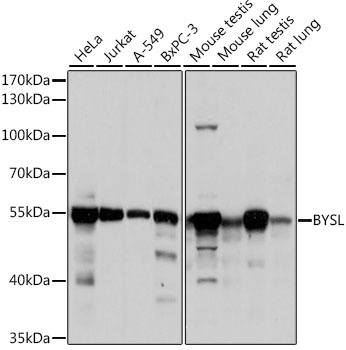
Western blot analysis of various lysates using BYSL Rabbit pAb (CAB11993) at 1:1000 dilution. Secondary antibody: HRP-conjugated Goat anti-Rabbit IgG (H+L) (CABS014) at 1:10000 dilution. Lysates/proteins: 25μg per lane. Blocking buffer: 3% nonfat dry milk in TBST. Detection: ECL Basic Kit (AbGn00020). Exposure time: 10s.
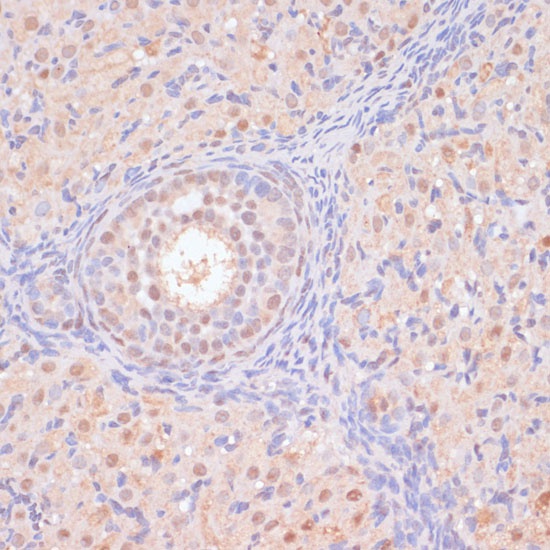
Immunohistochemistry analysis of paraffin-embedded Rat ovary using BYSL Rabbit pAb (CAB11993) at dilution of 1:100 (40x lens). Microwave antigen retrieval performed with 0.01M PBS Buffer (pH 7.2) prior to IHC staining.
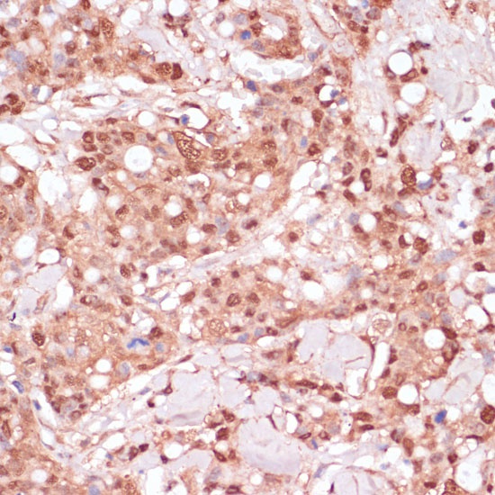
Immunohistochemistry analysis of paraffin-embedded Human breast cancer using BYSL Rabbit pAb (CAB11993) at dilution of 1:100 (40x lens). Microwave antigen retrieval performed with 0.01M PBS Buffer (pH 7.2) prior to IHC staining.