The C1QA Antibody (CAB1821) is a high-quality antibody developed for reliable detection and analysis of target proteins. C1QA is a subunit of the C1q protein, which is involved in the classical pathway of the complement system and plays a crucial role in the recognition and clearance of pathogens and damaged cells.Raised in rabbits, this antibody is highly specific to human samples and has been validated for use in Western blot applications. By binding to the C1QA protein, the antibody enables precise detection and analysis in a variety of cell types, making it an essential component for studies in immunology and infectious diseases.
This antibody is validated for use in WB, ELISA applications and has demonstrated reactivity against Human, Mouse, Rat samples.
Product Name:
C1QA Antibody
SKU:
CAB1821
Size:
20μL, 100μL
Reactivity:
Human, Mouse, Rat
Conjugate:
Unconjugated
Immunogen:
Recombinant protein (or fragment).This information is considered to be commercially sensitive.
Recommended starting concentration is 1 μg/mL. Please optimize the concentration based on your specific assay requirements.
Synonyms:
C1QD1, C1QA
Positive Sample:
RPMI 8226
Cellular Localization:
Secreted.
Calculated MW:
26kDa
Observed MW:
30kDa
This gene encodes the A-chain polypeptide of serum complement subcomponent C1q, which associates with C1r and C1s to yield the first component of the serum complement system. C1q deficiency is associated with lupus erythematosus and glomerulonephritis. C1q is composed of 18 polypeptide chains which include 6 A-chains, 6 B-chains, and 6 C-chains. Each chain contains an N-terminal collagen-like region and a C-terminal C1q globular domain. Alternate splicing results in multiple transcript variants.
Purification Method
Affinity purification
Gene ID
712
RRID
AB_2763858
Buffer Information
Store at -20℃. Avoid freeze / thaw cycles. Buffer: PBS containing 50% glycerol, preserved with proclin300 or sodium azide, pH 7.3.
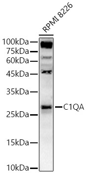
Western blot analysis of lysates from RPMI 8226 cells, using C1QA Rabbit pAb (CAB1821) at 1:2000 dilution. Secondary antibody: HRP-conjugated Goat anti-Rabbit IgG (H+L) (CABS014) at 1:10000 dilution. Lysates/proteins: 25μg per lane. Blocking buffer: 3% nonfat dry milk in TBST. Detection: ECL Basic Kit (AbGn00020). Exposure time: 30s.