The CAB39 Antibody (CAB16125) is a high-quality antibody developed for reliable detection and analysis of target proteins. CAB39, also known as MO25, is a crucial component of the LKB1-STRAD-MO25 complex, which plays a key role in regulating cell growth, metabolism, and polarity. This antibody, produced in rabbits, is highly specific to human samples and has been validated for use in Western blot applications.By targeting the CAB39 protein, this antibody enables researchers to detect and analyze the expression levels and localization of CAB39 in various cell types.
This antibody is validated for use in WB, IF/ICC, ELISA applications and has demonstrated reactivity against Human, Mouse, Rat samples.
Product Name:
CAB39 Antibody
SKU:
CAB16125
Size:
20μL, 100μL
Reactivity:
Human, Mouse, Rat
Conjugate:
Unconjugated
Immunogen:
Synthetic peptide. This information is considered to be commercially sensitive.
Recommended starting concentration is 1 μg/mL. Please optimize the concentration based on your specific assay requirements.
Synonyms:
MO25, CGI-66, CAB39
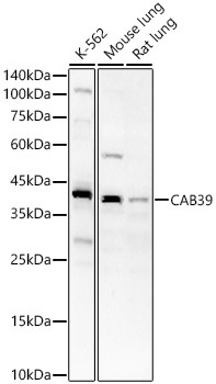
Positive Sample:
K-562, Mouse lung, Rat lung
Cellular Localization:
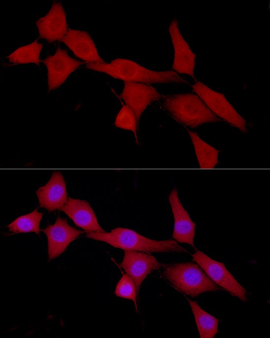
Cytoplasm.
Calculated MW:
40kDa
Observed MW:
39kDa
Enables kinase binding activity and protein serine/threonine kinase activator activity. Involved in intracellular signal transduction; peptidyl-serine phosphorylation; and positive regulation of protein phosphorylation. Located in extracellular exosome. Implicated in hepatocellular carcinoma. Biomarker of hepatocellular carcinoma and pancreatic cancer.
Purification Method
Affinity purification
Gene ID
51719
RRID
AB_2763570
Buffer Information
Store at -20℃. Avoid freeze / thaw cycles. Buffer: PBS containing 50% glycerol, preserved with proclin300 or sodium azide, pH 7.3.
Western blot analysis of various lysates, using CAB39 Rabbit pAb (CAB16125) at 1:700 dilution. Secondary antibody: HRP-conjugated Goat anti-Rabbit IgG (H+L) (CABS014) at 1:10000 dilution. Lysates/proteins: 25μg per lane. Blocking buffer: 3% nonfat dry milk in TBST. Detection: ECL Basic Kit (AbGn00020). Exposure time: 60s.
Immunofluorescence analysis of NIH/3T3 cells using CAB39 Rabbit pAb (CAB16125) at dilution of 1:100 (40x lens). Secondary antibody: Cy3-conjugated Goat anti-Rabbit IgG (H+L) (CABS007) at 1:500 dilution. Blue: DAPI for nuclear staining.