The CAD Monoclonal Antibody (CAB6849) is a high-quality antibody developed for reliable detection and analysis of target proteins. The antibody, raised in rabbits, is highly specific to human CAD and is validated for use in various applications such as Western blot and immunohistochemistry.CAD, also known as carbamoyl-phosphate synthetase 2, aspartate transcarbamylase, and dihydroorotase, plays a crucial role in de novo pyrimidine biosynthesis. Dysregulation of CAD expression or activity has been implicated in various diseases, including cancer and autoimmune disorders.
This antibody is validated for use in WB, IF/ICC, ELISA applications and has demonstrated reactivity against Human samples.
Product Name:
CAD Monoclonal Antibody
SKU:
CAB6849
Size:
20μL, 100μL
Reactivity:
Human
Clone Number:
ARC1426
Conjugate:
Unconjugated
Immunogen:
Synthetic peptide. This information is considered to be commercially sensitive.
The de novo synthesis of pyrimidine nucleotides is required for mammalian cells to proliferate. This gene encodes a trifunctional protein which is associated with the enzymatic activities of the first 3 enzymes in the 6-step pathway of pyrimidine biosynthesis: carbamoylphosphate synthetase (CPS II), aspartate transcarbamoylase, and dihydroorotase. This protein is regulated by the mitogen-activated protein kinase (MAPK) cascade, which indicates a direct link between activation of the MAPK cascade and de novo biosynthesis of pyrimidine nucleotides. Alternative splicing results in multiple transcript variants encoding different isoforms.
Purification Method
Affinity purification
Gene ID
790
Buffer Information
Store at -20℃. Avoid freeze / thaw cycles. Buffer: PBS containing 50% glycerol and 0.05% BSA, preserved with proclin300 or sodium azide, pH 7.3.
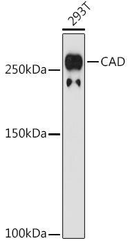
Western blot analysis of lysates from 293T cells, using CAD Rabbit mAb (CAB6849) at 1:1000 dilution. Secondary antibody: HRP-conjugated Goat anti-Rabbit IgG (H+L) (CABS014) at 1:10000 dilution. Lysates/proteins: 25μg per lane. Blocking buffer: 3% nonfat dry milk in TBST. Detection: ECL Basic Kit (AbGn00020). Exposure time: 180s.
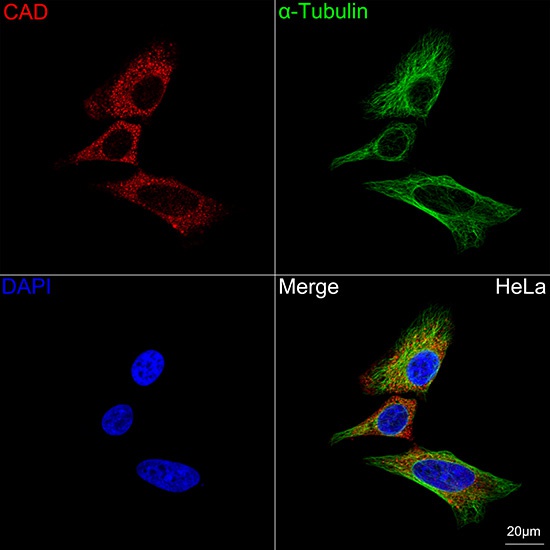
Confocal imaging of HeLa cells using CAD Rabbit mAb (CAB6849, dilution 1:100) followed by a further incubation with Cy3 Goat Anti-Rabbit IgG (H+L) (CABS007, dilution 1:500) (Red). The cells were counterstained with α-Tubulin Mouse mAb (AC012, dilution 1:400) followed by incubation with ABflo® 488-conjugated Goat Anti-Mouse IgG (H+L) Ab (CABS076, dilution 1:500) (Green). DAPI was used for nuclear staining (Blue). Objective: 100x.