The CADM1 Antibody (CAB1892) is a high-quality antibody developed for reliable detection and analysis of target proteins. Enables signaling receptor binding activity. Involved in several processes, including cell recognition; positive regulation of cytokine production; and susceptibility to natural killer cell mediated cytotoxicity. Located in plasma membrane. Implicated in breast carcinoma and prostate cancer. Biomarker of cervix uteri carcinoma in situ.
This antibody is validated for use in WB, ELISA applications and has demonstrated reactivity against Human, Mouse, Rat samples.
Product Name:
CADM1 Antibody
SKU:
CAB1892
Size:
100μL, 20μL
Reactivity:
Human, Mouse, Rat
Conjugate:
Unconjugated
Immunogen:
Recombinant protein (or fragment).This information is considered to be commercially sensitive.
Tested Applications:
WBELISA
Recommended Dilution:
WB
1:500 - 1:1000
ELISA
Recommended starting concentration is 1 μg/mL. Please optimize the concentration based on your specific assay requirements.
Cell Junction, Cell Membrane, Single-Pass Type I Membrane Protein, Synapse.
Calculated MW:
49kDa
Observed MW:
50kDa
Enables signaling receptor binding activity. Involved in several processes, including cell recognition; positive regulation of cytokine production; and susceptibility to natural killer cell mediated cytotoxicity. Located in plasma membrane. Implicated in breast carcinoma and prostate cancer. Biomarker of cervix uteri carcinoma in situ.
Purification Method
Affinity purification
Gene ID
23705
RRID
AB_2763924
Buffer Information
Store at -20℃. Avoid freeze / thaw cycles. Buffer: PBS containing 50% glycerol, preserved with proclin300 or sodium azide, pH 7.3.
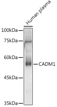
Western blot analysis of lysates from Human plasma, using CADM1 Rabbit pAb (CAB1892) at 1:1000 dilution. Secondary antibody: HRP-conjugated Goat anti-Rabbit IgG (H+L) (AS014) at 1:10000 dilution. Lysates/proteins: 25μg per lane. Blocking buffer: 3% nonfat dry milk in TBST. Detection: ECL Basic Kit (AbGn00020). Exposure time: 180s.
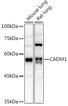
Western blot analysis of various lysates using CADM1 Rabbit pAb (CAB1892) at 1:1000 dilution. Secondary antibody: HRP-conjugated Goat anti-Rabbit IgG (H+L) (AS014) at 1:10000 dilution. Lysates/proteins: 25μg per lane. Blocking buffer: 3% nonfat dry milk in TBST. Detection: ECL Enhanced Kit (AbGn00021). Exposure time: 180s.