The Calponin Monoclonal Antibody (CAB3734) is a high-quality antibody developed for reliable detection and analysis of target proteins. This antibody, generated in rabbits, exhibits high specificity and sensitivity towards human samples, making it ideal for use in various research applications, such as immunohistochemistry and immunofluorescence.Calponin is a key regulator of actin cytoskeleton dynamics and plays a crucial role in cell migration, adhesion, and contraction. Dysregulation of calponin expression has been linked to a variety of diseases, including cardiovascular disorders and cancer.
This antibody is validated for use in WB, IHC-P, ELISA, IF-P applications and has demonstrated reactivity against Human, Mouse, Rat samples.
Product Name:
Calponin Monoclonal Antibody
SKU:
CAB3734
Size:
20μL, 100μL
Reactivity:
Human, Mouse, Rat
Clone Number:
ARC0232
Conjugate:
Unconjugated
Immunogen:
Synthetic peptide. This information is considered to be commercially sensitive.
Recommended starting concentration is 1 μg/mL. Please optimize the concentration based on your specific assay requirements.
Synonyms:
SMCC, Sm-Calp, HEL-S-14, Calponin
Positive Sample:
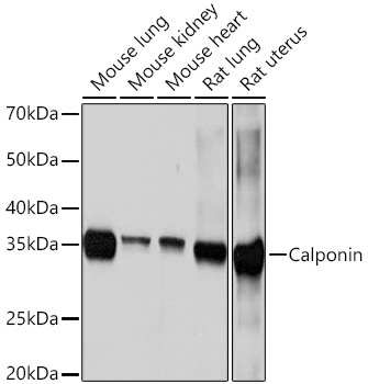
Mouse lung, Mouse kidney, Mouse heart, Rat lung, Rat uterus
Cellular Localization:
Cytoskeleton, Focal Adhesion.
Calculated MW:
33kDa
Observed MW:
35kDa
Predicted to enable actin binding activity. Involved in negative regulation of vascular associated smooth muscle cell proliferation. Located in cytoskeleton.
Purification Method
Affinity purification
Gene ID
1264
RRID
AB_2863131
Buffer Information
Store at -20℃. Avoid freeze / thaw cycles. Buffer: PBS containing 50% glycerol and 0.05% BSA, preserved with proclin300 or sodium azide, pH 7.3.
Western blot analysis of various lysates using Calponin Rabbit mAb (CAB3734) at 1:1000 dilution. Secondary antibody: HRP-conjugated Goat anti-Rabbit IgG (H+L) (CABS014) at 1:10000 dilution. Lysates/proteins: 25μg per lane. Blocking buffer: 3% nonfat dry milk in TBST. Detection: ECL Basic Kit (AbGn00020). Exposure time: 10s.
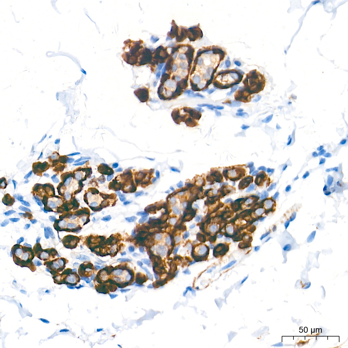
Immunohistochemistry analysis of paraffin-embedded Human breast tissue using Calponin Rabbit mAb (CAB3734) at a dilution of 1:1000 (40x lens). High pressure antigen retrieval performed with 0.01M Tris-EDTA Buffer (pH 9.0) prior to IHC staining.
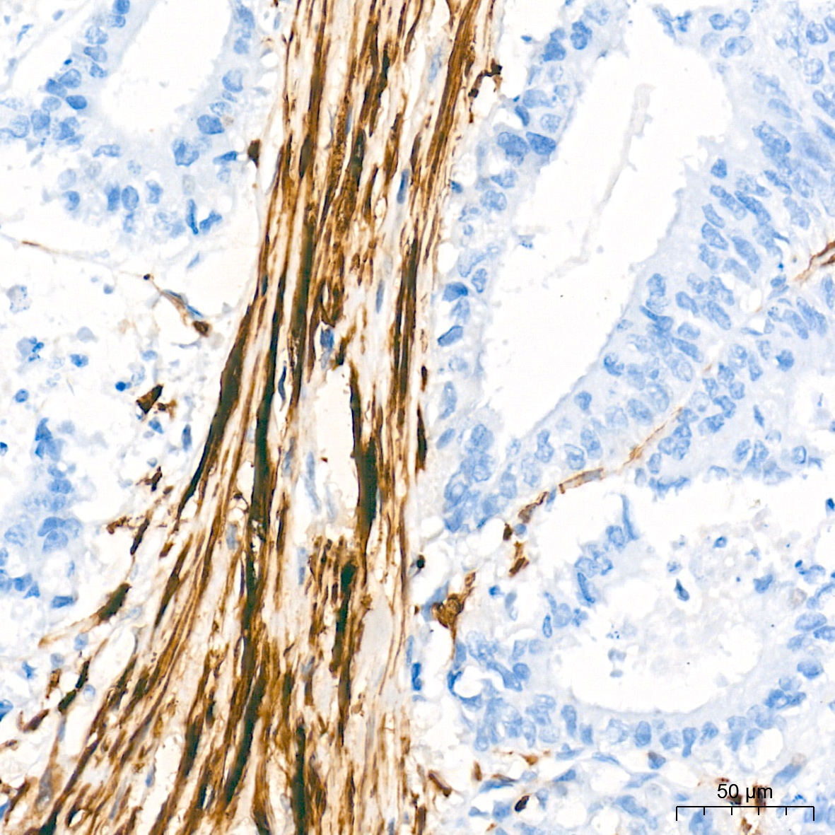
Immunohistochemistry analysis of paraffin-embedded Human colon carcinoma tissue using Calponin Rabbit mAb (CAB3734) at a dilution of 1:1000 (40x lens). High pressure antigen retrieval performed with 0.01M Tris-EDTA Buffer (pH 9.0) prior to IHC staining.
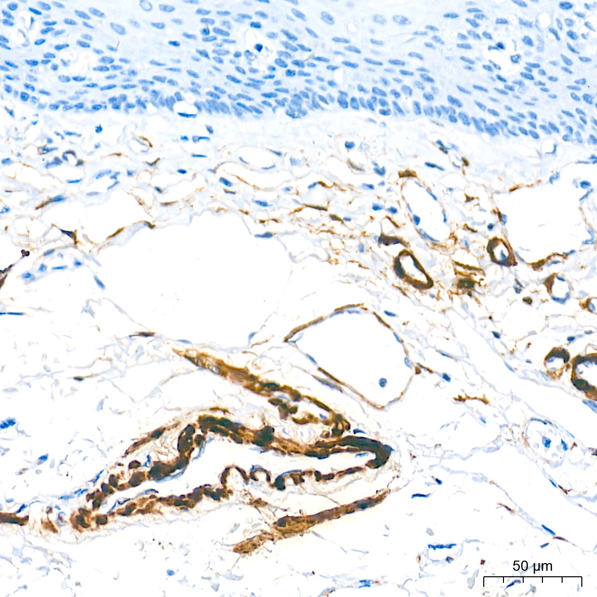
Immunohistochemistry analysis of paraffin-embedded Human esophagus tissue using Calponin Rabbit mAb (CAB3734) at a dilution of 1:1000 (40x lens). High pressure antigen retrieval performed with 0.01M Tris-EDTA Buffer (pH 9.0) prior to IHC staining.
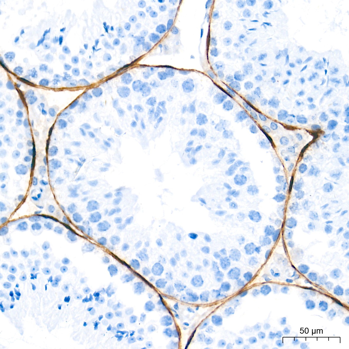
Immunohistochemistry analysis of paraffin-embedded Mouse testis tissue using Calponin Rabbit mAb (CAB3734) at a dilution of 1:1000 (40x lens). High pressure antigen retrieval performed with 0.01M Tris-EDTA Buffer (pH 9.0) prior to IHC staining.
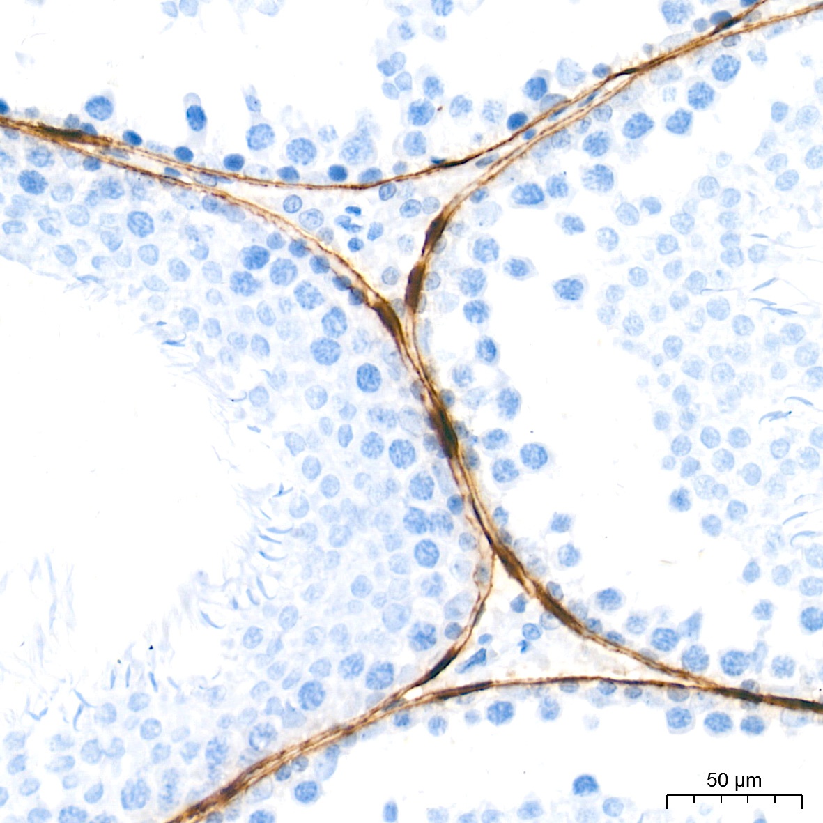
Immunohistochemistry analysis of paraffin-embedded Rat testis tissue using Calponin Rabbit mAb (CAB3734) at a dilution of 1:1000 (40x lens). High pressure antigen retrieval performed with 0.01M Tris-EDTA Buffer (pH 9.0) prior to IHC staining.
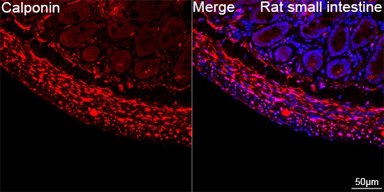
Confocal imaging of paraffin-embedded Rat small intestine tissue using Calponin Rabbit mAb (CAB3734, dilution 1:200) followed by a further incubation with Cy3 Goat Anti-Rabbit IgG (H+L) (CABS007, dilution 1:500) (Red). DAPI was used for nuclear staining (Blue). Objective: 40x. Perform high pressure antigen retrieval with 0.01 M citrate buffer (pH 6.0) prior to IF staining.