The Caspase-3 p12 Monoclonal Antibody (CAB19664) is a high-quality antibody developed for reliable detection and analysis of target proteins. Caspase-3 is a key player in the execution phase of apoptosis, cleaving various cellular substrates and ultimately leading to cell death. This antibody, raised in rabbits, is specifically designed to target the p12 subunit of Caspase-3 and is highly reactive with human samples.Validated for use in Western blot applications, this antibody enables precise detection and analysis of Caspase-3 in various cell types and tissues. Its high specificity ensures reliable and accurate results for researchers studying cell death pathways, cancer biology, and neurodegenerative diseases where dysregulation of apoptosis is a common feature.
This antibody is validated for use in WB, IHC-P, ELISA applications and has demonstrated reactivity against Human, Mouse, Rat samples.
Product Name:
Caspase-3 p12 Monoclonal Antibody
SKU:
CAB19664
Size:
20μL, 100μL
Reactivity:
Human, Mouse, Rat
Clone Number:
ARC0143
Conjugate:
Unconjugated
Immunogen:
Recombinant protein (or fragment).This information is considered to be commercially sensitive.
Recommended starting concentration is 1 μg/mL. Please optimize the concentration based on your specific assay requirements.
Synonyms:
CPP32, SCA-1, CPP32B
Positive Sample:
Jurkat treated with Etoposide, NIH/3T3 treated with Etoposide
Cellular Localization:
Cytoplasm.
Calculated MW:
32kDa
Observed MW:
12kDa/32kDa
The protein encoded by this gene is a cysteine-aspartic acid protease that plays a central role in the execution-phase of cell apoptosis. The encoded protein cleaves and inactivates poly(ADP-ribose) polymerase while it cleaves and activates sterol regulatory element binding proteins as well as caspases 6, 7, and 9. This protein itself is processed by caspases 8, 9, and 10. It is the predominant caspase involved in the cleavage of amyloid-beta 4A precursor protein, which is associated with neuronal death in Alzheimer's disease.
Purification Method
Affinity purification
Gene ID
836
Buffer Information
Store at -20℃. Avoid freeze / thaw cycles. Buffer: PBS containing 50% glycerol and 0.05% BSA, preserved with proclin300 or sodium azide, pH 7.3.
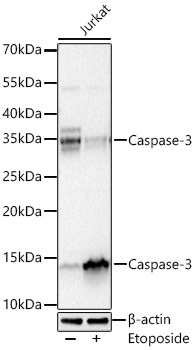
Western blot analysis of lysates from Jurkat cells using Caspase-3 Rabbit mAb (CAB19664) at 1:5000 dilution incubated overnight at 4℃. Jurkat cells were treated with Etoposide (25 μM) at 37℃ for 5 hours. Secondary antibody: HRP-conjugated Goat anti-Rabbit IgG (H+L) (CABS014) at 1:10000 dilution. Lysates/proteins: 30 μg per lane. Blocking buffer: 3 % nonfat dry milk in TBST. Detection: ECL Basic Kit (AbGn00020). Exposure time: 20s.
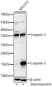
Western blot analysis of lysates from NIH/3T3 cells using Caspase-3 Rabbit mAb (CAB19664) at 1:5000 dilution incubated overnight at 4℃. NIH/3T3 cells were treated with Stauroposide (1 μM) at 37℃ for 5 hours. Secondary antibody: HRP-conjugated Goat anti-Rabbit IgG (H+L) (CABS014) at 1:10000 dilution. Lysates/proteins: 30 μg per lane. Blocking buffer: 3 % nonfat dry milk in TBST. Detection: ECL Basic Kit (AbGn00020). Exposure time: 45s.
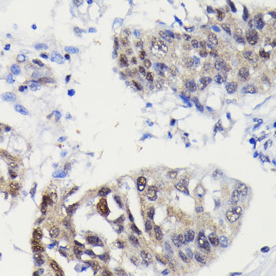
Immunohistochemistry analysis of paraffin-embedded Human lung cancer using Caspase-3 Rabbit mAb (CAB19664) at dilution of 1:100 (40x lens). Microwave antigen retrieval performed with 0.01M PBS Buffer (pH 7.2) prior to IHC staining.