The Cathepsin B Antibody (CAB0967) is a high-quality antibody developed for reliable detection and analysis of target proteins. This antibody, generated in rabbits, shows high specificity and sensitivity towards human samples, making it suitable for use in Western blot experiments.Cathepsin B is a key player in cancer progression, metastasis, and invasion, making it a potential therapeutic target for cancer treatment.
This antibody is validated for use in WB, IHC-P, IF/ICC, ELISA applications and has demonstrated reactivity against Human, Mouse, Rat samples.
Product Name:
Cathepsin B Antibody
SKU:
CAB0967
Size:
20μL, 100μL
Reactivity:
Human, Mouse, Rat
Conjugate:
Unconjugated
Immunogen:
Recombinant protein (or fragment).This information is considered to be commercially sensitive.
This gene encodes a member of the C1 family of peptidases. Alternative splicing of this gene results in multiple transcript variants. At least one of these variants encodes a preproprotein that is proteolytically processed to generate multiple protein products. These products include the cathepsin B light and heavy chains, which can dimerize to form the double chain form of the enzyme. This enzyme is a lysosomal cysteine protease with both endopeptidase and exopeptidase activity that may play a role in protein turnover. It is also known as amyloid precursor protein secretase and is involved in the proteolytic processing of amyloid precursor protein (APP). Incomplete proteolytic processing of APP has been suggested to be a causative factor in Alzheimer's disease, the most common cause of dementia. Overexpression of the encoded protein has been associated with esophageal adenocarcinoma and other tumors. Both Cathepsin B and Cathepsin L are involved in the cleavage of the spike protein from the severe acute respiratory syndrome coronavirus 2 (SARS-CoV-2) upon its entry to the human host cell. Multiple pseudogenes of this gene have been identified.
Purification Method
Affinity purification
Gene ID
1508
RRID
AB_2757486
Buffer Information
Store at -20℃. Avoid freeze / thaw cycles. Buffer: PBS containing 50% glycerol, preserved with proclin300 or sodium azide, pH 7.3.
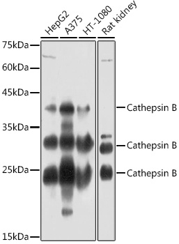
Western blot analysis of various lysates using Cathepsin B Rabbit pAb (CAB0967) at 1:1000 dilution. Secondary antibody: HRP-conjugated Goat anti-Rabbit IgG (H+L) (CABS014) at 1:10000 dilution. Lysates/proteins: 25μg per lane. Blocking buffer: 3% nonfat dry milk in TBST. Detection: ECL Basic Kit (AbGn00020). Exposure time: 60s.
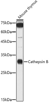
Western blot analysis of lysates from Mouse thymus, using Cathepsin B Rabbit pAb (CAB0967) at 1:1000 dilution. Secondary antibody: HRP-conjugated Goat anti-Rabbit IgG (H+L) (CABS014) at 1:10000 dilution. Lysates/proteins: 25μg per lane. Blocking buffer: 3% nonfat dry milk in TBST. Detection: ECL Enhanced Kit (AbGn00021). Exposure time: 180s.
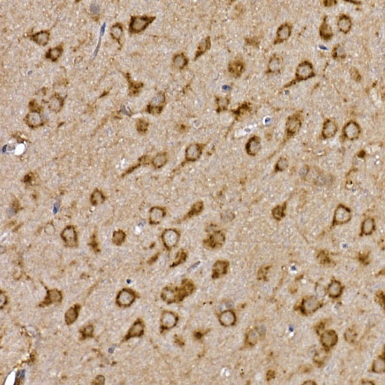
Immunohistochemistry analysis of paraffin-embedded Rat brain using Cathepsin B Rabbit pAb (CAB0967) at dilution of 1:100 (40x lens). High pressure antigen retrieval performed with 0.01M Citrate buffer (pH 6.0) prior to IHC staining.
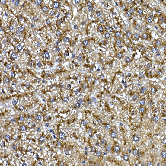
Immunohistochemistry analysis of paraffin-embedded Rat liver using Cathepsin B Rabbit pAb (CAB0967) at dilution of 1:100 (40x lens). High pressure antigen retrieval performed with 0.01M Citrate buffer (pH 6.0) prior to IHC staining.
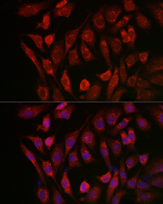
Immunofluorescence analysis of NIH/3T3 cells using Cathepsin B Rabbit pAb (CAB0967) at dilution of 1:100 (40x lens). Secondary antibody: Cy3-conjugated Goat anti-Rabbit IgG (H+L) (CABS007) at 1:500 dilution. Blue: DAPI for nuclear staining.