The Cathepsin D Monoclonal Antibody (CAB19680) is a high-quality antibody developed for reliable detection and analysis of target proteins. This antibody, generated in rabbits, exhibits strong reactivity towards human samples and has been validated for use in various applications such as Western blotting.Cathepsin D is a key enzyme involved in lysosomal function and is known to play a critical role in various physiological processes, including cell growth, differentiation, and apoptosis. Dysregulation of Cathepsin D has been implicated in several diseases, making it a promising target for research in fields such as cancer biology, neurodegenerative disorders, and metabolic diseases.
This antibody is validated for use in WB, IHC-P, ELISA applications and has demonstrated reactivity against Human, Mouse samples.
Product Name:
Cathepsin D Monoclonal Antibody
SKU:
CAB19680
Size:
20μL, 100μL
Reactivity:
Human, Mouse
Clone Number:
ARC0160
Conjugate:
Unconjugated
Immunogen:
Synthetic peptide. This information is considered to be commercially sensitive.
This gene encodes a member of the A1 family of peptidases. The encoded preproprotein is proteolytically processed to generate multiple protein products. These products include the cathepsin D light and heavy chains, which heterodimerize to form the mature enzyme. This enzyme exhibits pepsin-like activity and plays a role in protein turnover and in the proteolytic activation of hormones and growth factors. Mutations in this gene play a causal role in neuronal ceroid lipofuscinosis-10 and may be involved in the pathogenesis of several other diseases, including breast cancer and possibly Alzheimer's disease.
Purification Method
Affinity purification
Gene ID
1509
RRID
AB_2862731
Buffer Information
Store at -20℃. Avoid freeze / thaw cycles. Buffer: PBS containing 50% glycerol and 0.05% BSA, preserved with proclin300 or sodium azide, pH 7.3.
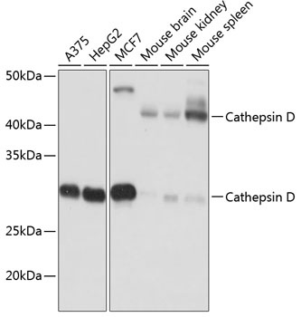
Western blot analysis of various lysates using Cathepsin D Rabbit mAb (CAB19680) at 1:1000 dilution. Secondary antibody: HRP-conjugated Goat anti-Rabbit IgG (H+L) (CABS014) at 1:10000 dilution. Lysates/proteins: 25μg per lane. Blocking buffer: 3% nonfat dry milk in TBST. Detection: ECL Basic Kit (AbGn00020). Exposure time: 10s.
Immunohistochemistry analysis of paraffin-embedded Human colon carcinoma tissue using Cathepsin D Rabbit mAb (CAB19680) at a dilution of 1:1000 (40x lens). High pressure antigen retrieval was performed with 0.01 M citrate buffer (pH 6.0) prior to IHC staining.
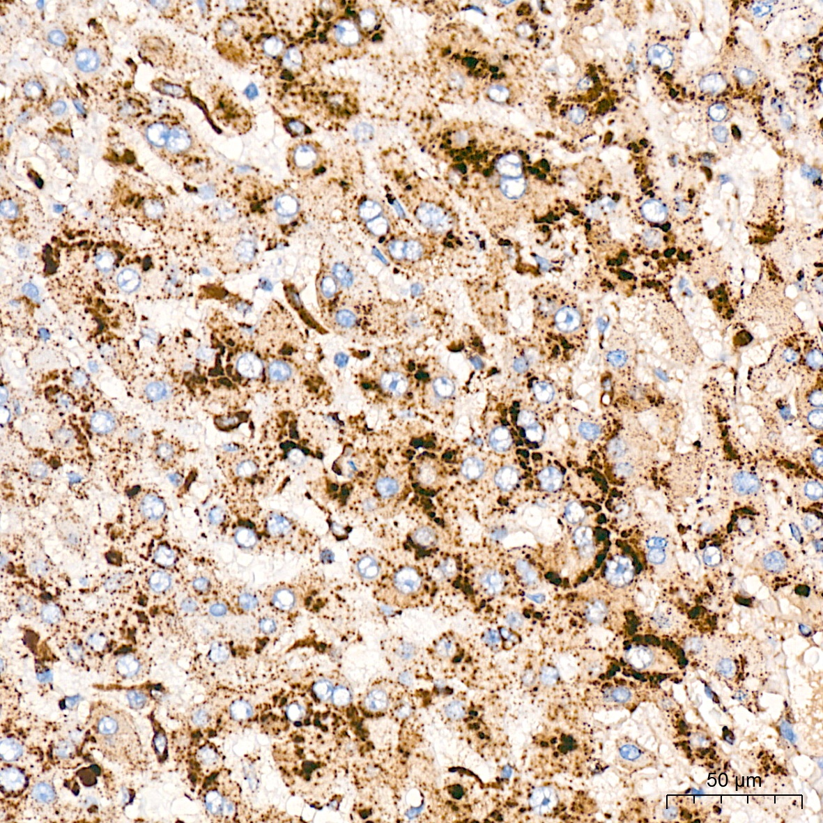
Immunohistochemistry analysis of paraffin-embedded Human liver tissue using Cathepsin D Rabbit mAb (CAB19680) at a dilution of 1:1000 (40x lens). High pressure antigen retrieval was performed with 0.01 M citrate buffer (pH 6.0) prior to IHC staining.
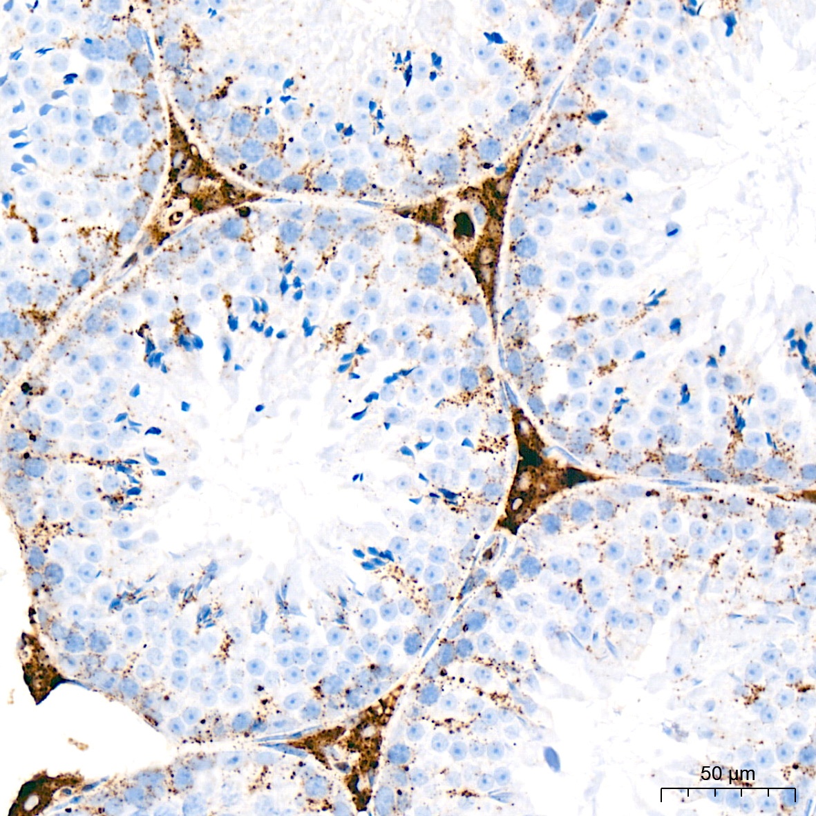
Immunohistochemistry analysis of paraffin-embedded Mouse testis tissue using Cathepsin D Rabbit mAb (CAB19680) at a dilution of 1:1000 (40x lens). High pressure antigen retrieval was performed with 0.01 M citrate buffer (pH 6.0) prior to IHC staining.
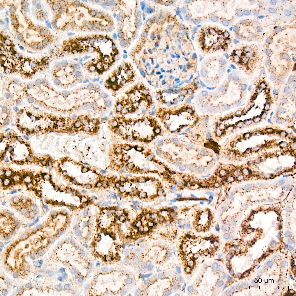
Immunohistochemistry analysis of paraffin-embedded Mouse kidney tissue using Cathepsin D Rabbit mAb (CAB19680) at a dilution of 1:1000 (40x lens). High pressure antigen retrieval was performed with 0.01 M citrate buffer (pH 6.0) prior to IHC staining.