The MCP-1 Antibody (CAB7277) is a high-quality antibody developed for reliable detection and analysis of target proteins. This gene is one of several cytokine genes clustered on the q-arm of chromosome 17. Chemokines are a superfamily of secreted proteins involved in immunoregulatory and inflammatory processes. The superfamily is divided into four subfamilies based on the arrangement of N-terminal cysteine residues of the mature peptide. This chemokine is a member of the CC subfamily which is characterized by two adjacent cysteine residues. This cytokine displays chemotactic activity for monocytes and basophils but not for neutrophils or eosinophils. It has been implicated in the pathogenesis of diseases characterized by monocytic infiltrates, like psoriasis, rheumatoid arthritis and atherosclerosis. It binds to chemokine receptors CCR2 and CCR4. Elevated expression of the encoded protein is associated with severe acute respiratory syndrome coronavirus 2 (SARS‐CoV‐2) infection. RRID AB_2767818 Gene ID 6347 Swiss Prot Synonym HC11; MCAF; MCP1; MCP-1; SCYA2; GDCF-2; SMC-CF; HSMCR30; CCL2/MCP-1
This antibody is validated for use in WB, IHC-P, ELISA applications and has demonstrated reactivity against Human, Mouse, Rat samples.
Product Name:
MCP-1 Antibody
SKU:
CAB7277
Size:
100μL, 20μL
Reactivity:
Human, Mouse, Rat
Clone Number:
-
Conjugate:
Unconjugated
Immunogen:
Recombinant protein (or fragment).This information is considered to be commercially sensitive.
Tested Applications:
WBIHC-PELISA
Recommended Dilution:
WB
1:1000 - 1:5000
IHC-P
1:50 - 1:200
ELISA
Recommended starting concentration is 1 μg/mL. Please optimize the concentration based on your specific assay requirements.
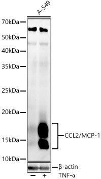
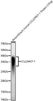
A-549 treated with TNF-α, Recombinant Human CCL2/MCP-1 Protein
Cellular Localization:
Secreted.
Calculated MW:
11kDa
Observed MW:
13-15kDa
This gene is one of several cytokine genes clustered on the q-arm of chromosome 17. Chemokines are a superfamily of secreted proteins involved in immunoregulatory and inflammatory processes. The superfamily is divided into four subfamilies based on the arrangement of N-terminal cysteine residues of the mature peptide. This chemokine is a member of the CC subfamily which is characterized by two adjacent cysteine residues. This cytokine displays chemotactic activity for monocytes and basophils but not for neutrophils or eosinophils. It has been implicated in the pathogenesis of diseases characterized by monocytic infiltrates, like psoriasis, rheumatoid arthritis and atherosclerosis. It binds to chemokine receptors CCR2 and CCR4. Elevated expression of the encoded protein is associated with severe acute respiratory syndrome coronavirus 2 (SARS‐CoV‐2) infection. RRID AB_2767818 Gene ID 6347 Swiss Prot Synonym HC11; MCAF; MCP1; MCP-1; SCYA2; GDCF-2; SMC-CF; HSMCR30; CCL2/MCP-1
Purification Method:
Affinity purification
Gene ID:
6347
RRID:
AB_2767818
Buffer Information:
Store at -20℃. Avoid freeze / thaw cycles. Buffer: PBS containing 50% glycerol, preserved with proclin300 or sodium azide, pH 7.3.
Western blot analysis of lysates from A-549 cells, using CCL2/MCP-1 Rabbit pAb (CAB7277) at 1:2000 dilution. A-549 cells were treated with TNF-α (20 ng/ml) at 37℃ for 30 minutes after serum-starvation overnight. Secondary antibody: HRP-conjugated Goat anti-Rabbit IgG (H+L) (AS014) at 1:10000 dilution. Lysates/proteins: 25μg per lane. Blocking buffer: 3% nonfat dry milk in TBST. Detection: ECL Enhanced Kit (AbGn00021). Exposure time: 30s.
Western blot analysis of Recombinant Human CCL2/MCP-1 Protein (RP01653), using CCL2/MCP-1 Rabbit pAb (CAB7277) at 1:2000 dilution. Secondary antibody: HRP-conjugated Goat anti-Rabbit IgG (H+L) (AS014) at 1:10000 dilution. Lysates/proteins: 10ng per lane. Blocking buffer: 3% nonfat dry milk in TBST. Detection: ECL Enhanced Kit (AbGn00021). Exposure time: 30s.
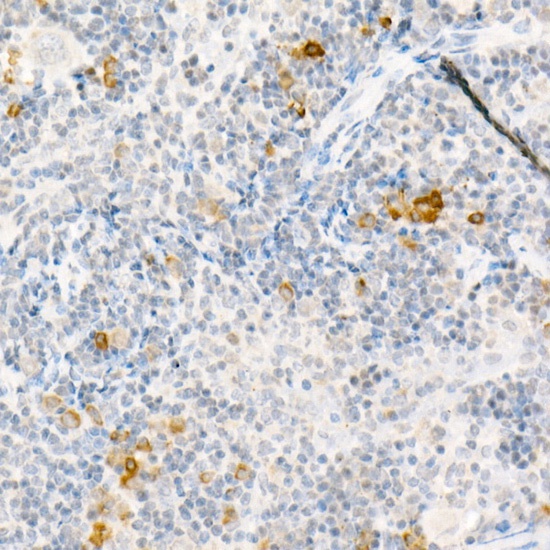
Immunohistochemistry analysis of paraffin-embedded Mouse spleen using CCL2/MCP-1 Rabbit pAb (CAB7277) at dilution of 1:50 (40x lens). High pressure antigen retrieval performed with 0.01M Citrate buffer (pH 6.0) prior to IHC staining.
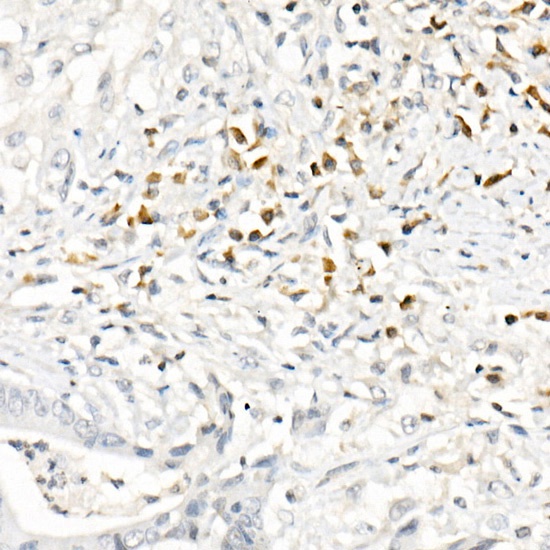
Immunohistochemistry analysis of paraffin-embedded Human colon carcinoma using CCL2/MCP-1 Rabbit pAb (CAB7277) at dilution of 1:50 (40x lens). High pressure antigen retrieval performed with 0.01M Citrate buffer (pH 6.0) prior to IHC staining.


")


