The Cyclin H Antibody (CAB0995) is a high-quality antibody developed for reliable detection and analysis of target proteins. This antibody, produced in rabbits, demonstrates high reactivity with human samples and is specifically validated for use in Western blot applications.Cyclin H is a key player in cell cycle progression, functioning as a regulatory subunit of the CDK7 kinase complex. It is essential for cell division and DNA replication, making it a crucial target for studies in cancer biology and developmental biology. With the ability to detect and analyze Cyclin H protein levels in various cell types, this antibody is well-suited for immunology and cancer research applications.
This antibody is validated for use in WB, IP, ELISA applications and has demonstrated reactivity against Human samples.
Product Name:
Cyclin H Antibody
SKU:
CAB0995
Size:
20μL, 100μL
Reactivity:
Human
Conjugate:
Unconjugated
Immunogen:
Recombinant protein (or fragment).This information is considered to be commercially sensitive.
The protein encoded by this gene belongs to the highly conserved cyclin family, whose members are characterized by a dramatic periodicity in protein abundance through the cell cycle. Cyclins function as regulators of CDK kinases. Different cyclins exhibit distinct expression and degradation patterns which contribute to the temporal coordination of each mitotic event. This cyclin forms a complex with CDK7 kinase and ring finger protein MAT1. The kinase complex is able to phosphorylate CDK2 and CDC2 kinases, thus functions as a CDK-activating kinase (CAK). This cyclin and its kinase partner are components of TFIIH, as well as RNA polymerase II protein complexes. They participate in two different transcriptional regulation processes, suggesting an important link between basal transcription control and the cell cycle machinery. A pseudogene of this gene is found on chromosome 4. Alternate splicing results in multiple transcript variants.
Purification Method
Affinity purification
Gene ID
902
RRID
AB_2757514
Buffer Information
Store at -20℃. Avoid freeze / thaw cycles. Buffer: PBS containing 50% glycerol, preserved with proclin300 or sodium azide, pH 7.3.
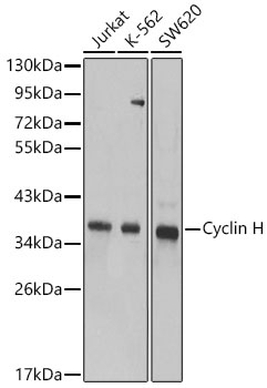
Western blot analysis of various lysates using Cyclin H Rabbit pAb (CAB0995) at 1:1000 dilution. Secondary antibody: HRP-conjugated Goat anti-Rabbit IgG (H+L) (CABS014) at 1:10000 dilution. Lysates / proteins: 25 μg per lane. Blocking buffer: 3 % nonfat dry milk in TBST. Detection: ECL Basic Kit (AbGn00020). Exposure time: 90s.
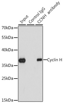
Immunoprecipitation analysis of 200 μg extracts of K562 cells using 1 μg Cyclin H antibody (CAB0995). Western blot was performed from the immunoprecipitate using Cyclin H antibody (CAB0995) at a dilution of 1:1000.