The CD106 Polyclonal Antibody (CAB23263) is a high-quality antibody developed for reliable detection and analysis of target proteins. This antibody is raised in rabbits and exhibits high reactivity with human samples, making it ideal for Western blot applications. By binding specifically to the CD106 protein, researchers can accurately detect and analyze its expression in various cell types, providing valuable insights into immunology and cancer research.
This antibody is validated for use in WB, ELISA applications and has demonstrated reactivity against Mouse samples.
Product Name:
CD106 Polyclonal Antibody
SKU:
CAB23263
Size:
20μL, 100μL
Reactivity:
Mouse
Conjugate:
Unconjugated
Immunogen:
Recombinant protein (or fragment).This information is considered to be commercially sensitive.
Tested Applications:
WBELISA
Recommended Dilution:
WB
1:500 - 1:1000
ELISA
Recommended starting concentration is 1 μg/mL. Please optimize the concentration based on your specific assay requirements.
Synonyms:
CD106, Vcam-1
Positive Sample:
RAW264.7(Negative control), Mouse spleen
Calculated MW:
81kDa
Observed MW:
85-100kDa
Predicted to enable integrin binding activity and primary amine oxidase activity. Acts upstream of or within several processes, including cellular response to glucose stimulus; chorio-allantoic fusion; and heterophilic cell-cell adhesion via plasma membrane cell adhesion molecules. Located in apical plasma membrane. Is expressed in several structures, including brain; embryo mesenchyme; extraembryonic component; heart; and hemolymphoid system. Human ortholog(s) of this gene implicated in hypertension. Orthologous to human VCAM1 (vascular cell adhesion molecule 1).
Purification Method
Affinity purification
Gene ID
22329
Buffer Information
Store at -20℃. Avoid freeze / thaw cycles. Buffer: PBS containing 50% glycerol, preserved with proclin300 or sodium azide, pH 7.3.
Western blot analysis of various lysates, using CD106 Rabbit pAb (CAB23263) at 1:2500 dilution. Secondary antibody: HRP-conjugated Goat anti-Rabbit IgG (H+L) (CABS014) at 1:10000 dilution. Lysates/proteins: 25μg per lane. Blocking buffer: 3% nonfat dry milk in TBST. Detection: ECL Basic Kit (AbGn00020). Exposure time: 30s.
 at 1:2500 dilution. Secondary antibody: HRP Goat Anti-Rabbit IgG (H+L) at 1:10000 dilution. Lysates/proteins: 25μg per lane. Blocking buffer: 3% nonfat dry milk in TBST.")
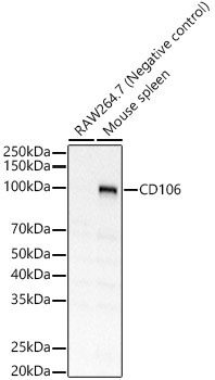
 at 1:2500 dilution. Secondary antibody: HRP Goat Anti-Rabbit IgG (H+L) at 1:10000 dilution. Lysates/proteins: 25μg per lane. Blocking buffer: 3% nonfat dry milk in TBST.")


![APC Anti-Mouse CD106 Antibody [M/K-2.7] (AGEL1174)](https://cdn11.bigcommerce.com/s-h68l9z2lnx/images/stencil/590x590/products/21705/606631/apc-anti-mouse-cd106-antibody-mk-2.7-agel1174__16617.1707498661.jpg?c=2 "APC Anti-Mouse CD106 Antibody [M/K-2.7] (AGEL1174)")
![APC Anti-Mouse CD106 Antibody [M/K-2.7] (AGEL1174)](https://cdn11.bigcommerce.com/s-h68l9z2lnx/images/stencil/590x590/products/21705/719941/AGEL1174_spectral__31788.1775232788.png?c=2 "APC Anti-Mouse CD106 Antibody [M/K-2.7] (AGEL1174)")
![Purified Anti-Mouse CD106 Antibody [M/K-2.7] (AGEL0334)](https://cdn11.bigcommerce.com/s-h68l9z2lnx/images/stencil/590x590/products/20207/605579/anti-mouse-cd106-antibody-mk-2.7-agel0334__99518.1707495305.jpg?c=2 "Purified Anti-Mouse CD106 Antibody [M/K-2.7] (AGEL0334)")
![Biotin Anti-Mouse CD106 Antibody [M/K-2.7] (AGEL0335)](https://cdn11.bigcommerce.com/s-h68l9z2lnx/images/stencil/590x590/products/20208/605693/biotin-anti-mouse-cd106-antibody-mk-2.7-agel0335__03402.1707495664.jpg?c=2 "Biotin Anti-Mouse CD106 Antibody [M/K-2.7] (AGEL0335)")