The CD1D Antibody (CAB1760) is a high-quality antibody developed for reliable detection and analysis of target proteins. This antibody, generated in rabbits, exhibits high reactivity with human samples and has been validated for use in Western blot applications. By specifically binding to the CD1d protein, researchers can effectively detect and analyze its expression in various cell types, making it an essential tool for studies in immunology and autoimmune diseases.
This antibody is validated for use in WB, IF/ICC, ELISA applications and has demonstrated reactivity against Human, Mouse samples.
Product Name:
CD1D Antibody
SKU:
CAB1760
Size:
20μL, 100μL
Reactivity:
Human, Mouse
Conjugate:
Unconjugated
Immunogen:
Recombinant protein (or fragment).This information is considered to be commercially sensitive.
Recommended starting concentration is 1 μg/mL. Please optimize the concentration based on your specific assay requirements.
Synonyms:
R3, CD1A, R3G1, CD1D
Positive Sample:
THP-1, Jurkat
Cellular Localization:
Cell Membrane, Endosome Membrane, Lysosome Membrane, Single-Pass Type I Membrane Protein.
Calculated MW:
38kDa
Observed MW:
38kDa/50kDa
This gene encodes a divergent member of the CD1 family of transmembrane glycoproteins, which are structurally related to the major histocompatibility complex (MHC) proteins and form heterodimers with beta-2-microglobulin. The CD1 proteins mediate the presentation of primarily lipid and glycolipid antigens of self or microbial origin to T cells. The human genome contains five CD1 family genes organized in a cluster on chromosome 1. The CD1 family members are thought to differ in their cellular localization and specificity for particular lipid ligands. The protein encoded by this gene localizes to late endosomes and lysosomes via a tyrosine-based motif in the cytoplasmic tail. Two transcript variants encoding different isoforms have been found for this gene.
Purification Method
Affinity purification
Gene ID
912
RRID
AB_2763803
Buffer Information
Store at -20℃. Avoid freeze / thaw cycles. Buffer: PBS containing 50% glycerol, preserved with proclin300 or sodium azide, pH 7.3.
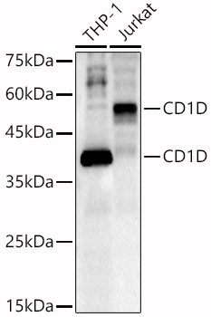
Western blot analysis of various lysates, using CD1D Rabbit pAb (CAB1760) at 1:1000 dilution. Secondary antibody: HRP-conjugated Goat anti-Rabbit IgG (H+L) (CABS014) at 1:10000 dilution. Lysates/proteins: 25μg per lane. Blocking buffer: 3% nonfat dry milk in TBST. Detection: ECL Basic Kit (AbGn00020). Exposure time: 60s.
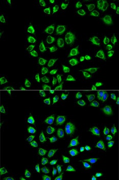
Immunofluorescence analysis of HeLa cells using CD1D Rabbit pAb (CAB1760). Secondary antibody: Cy3-conjugated Goat anti-Rabbit IgG (H+L) (CABS007) at 1:500 dilution. Blue: DAPI for nuclear staining.