The CD209 Monoclonal Antibody (CAB9649) is a high-quality antibody developed for reliable detection and analysis of target proteins. This antibody, developed using rabbit monoclonal technology, exhibits high specificity and sensitivity in detecting CD209 in human samples. Validated for use in various applications such as Western blotting, the antibody binds specifically to the CD209 protein, enabling accurate and reliable detection in a wide range of cell types.CD209, also known as DC-SIGN, is a key player in the immune system, particularly in the recognition and uptake of pathogens by dendritic cells. Its ability to interact with a variety of pathogens, including viruses, bacteria, and parasites, makes it a valuable target for research in infectious diseases and vaccine development.
This antibody is validated for use in WB, IHC-P, IF/ICC, ELISA applications and has demonstrated reactivity against Human, Mouse, Rat samples.
Product Name:
CD209 Monoclonal Antibody
SKU:
CAB9649
Size:
20μL, 100μL
Reactivity:
Human, Mouse, Rat
Clone Number:
ARC1679
Conjugate:
Unconjugated
Immunogen:
Synthetic peptide. This information is considered to be commercially sensitive.
Cell Membrane, Single-Pass Type Ii Membrane Protein, Secreted.
Calculated MW:
46kDa
Observed MW:
45-55kDa
This gene encodes a C-type lectin that functions in cell adhesion and pathogen recognition. This receptor recognizes a wide range of evolutionarily divergent pathogens with a large impact on public health, including leprosy and tuberculosis mycobacteria, the Ebola, hepatitis C, HIV-1 and Dengue viruses, and the SARS-CoV acute respiratory syndrome coronavirus. The protein is organized into four distinct domains: a C-terminal carbohydrate recognition domain, a flexible tandem-repeat neck domain, a transmembrane region and an N-terminal cytoplasmic domain involved in internalization. This gene is closely related in terms of both sequence and function to a neighboring gene, CLEC4M (Gene ID: 10332), also known as L-SIGN. The two genes differ in viral recognition and expression patterns, with this gene showing high expression on the surface of dendritic cells. Polymorphisms in the neck region are associated with protection from HIV-1 infection, while single nucleotide polymorphisms in the promoter of this gene are associated with differing resistance and susceptibility to and severity of infectious disease, including rs4804803, which is associated with SARS severity.
Purification Method
Affinity purification
Gene ID
30835
RRID
AB_2863747
Buffer Information
Store at -20℃. Avoid freeze / thaw cycles. Buffer: PBS containing 50% glycerol and 0.05% BSA, preserved with proclin300 or sodium azide, pH 7.3.
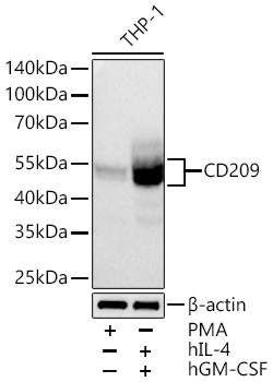
Western blot analysis of lysates from THP-1 cells using CD209 Rabbit mAb (CAB9649) at 1:1000 dilution incubated overnight at 4℃. THP-1 cells were treated with PMA (80nM) for 72 hours, or treated with hIL-4 (20 ng/mL) for 4 days and hGM-CSF (40 ng/mL) for 4 days. Secondary antibody: HRP-conjugated Goat anti-Rabbit IgG (H+L) (CABS014) at 1:10000 dilution. Lysates/proteins: 30 μg per lane. Blocking buffer: 3% nonfat dry milk in TBST. Detection: ECL Basic Kit (AbGn00020). Exposure time: 60 s.
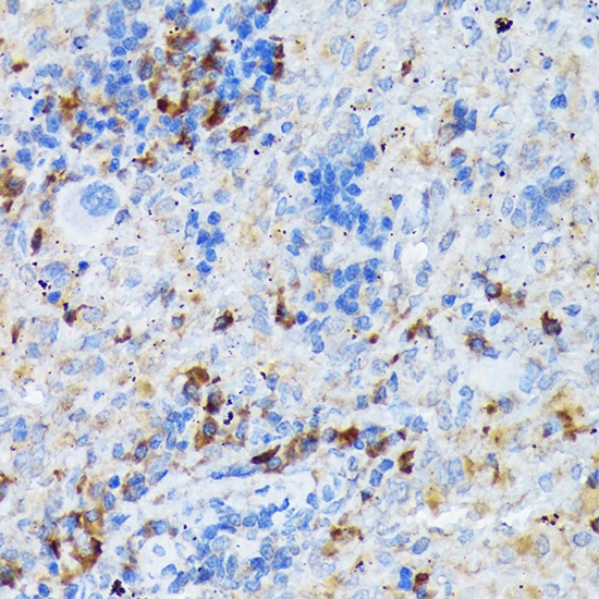
Immunohistochemistry analysis of paraffin-embedded Rat spleen using CD209 Rabbit mAb (CAB9649) at dilution of 1:100 (40x lens). Microwave antigen retrieval performed with 0.01M Tris/EDTA Buffer (pH 9.0) prior to IHC staining.
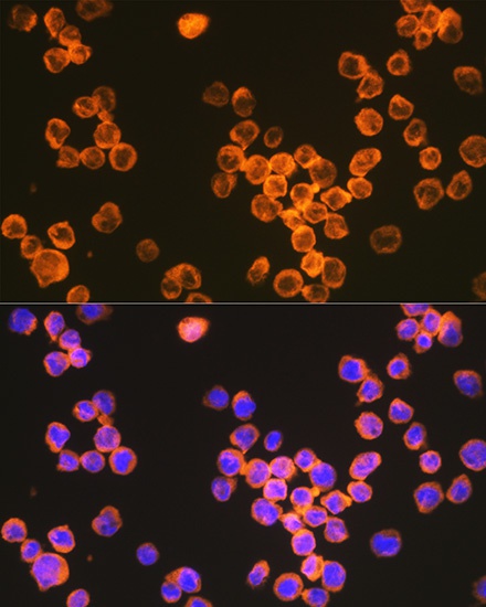
Immunofluorescence analysis of THP-1 cells using CD209 Rabbit mAb (CAB9649) at dilution of 1:100 (40x lens). Secondary antibody: Cy3-conjugated Goat anti-Rabbit IgG (H+L) (CABS007) at 1:500 dilution. Blue: DAPI for nuclear staining.