The CD3H Antibody (CAB2058) is a high-quality antibody developed for reliable detection and analysis of target proteins. This antibody, produced in rabbits, exhibits high reactivity with human samples and has been validated for use in Western blot applications. By binding specifically to the CD247 protein, this antibody enables precise detection and analysis in various cell types, making it an invaluable resource for studies in immunology and cancer research.CD247, also known as the CD3 zeta chain, is a critical component of the T cell receptor complex and is essential for signal transduction during T cell activation.
This antibody is validated for use in WB, IHC-P, IF/ICC, ELISA applications and has demonstrated reactivity against Human, Mouse, Rat samples.
Product Name:
CD3H Antibody
SKU:
CAB2058
Size:
20μL, 100μL
Reactivity:
Human, Mouse, Rat
Conjugate:
Unconjugated
Immunogen:
Recombinant protein (or fragment).This information is considered to be commercially sensitive.
The protein encoded by this gene is T-cell receptor zeta, which together with T-cell receptor alpha/beta and gamma/delta heterodimers, and with CD3-gamma, -delta and -epsilon, forms the T-cell receptor-CD3 complex. The zeta chain plays an important role in coupling antigen recognition to several intracellular signal-transduction pathways. Low expression of the antigen results in impaired immune response. Two alternatively spliced transcript variants encoding distinct isoforms have been found for this gene.
Purification Method
Affinity purification
Gene ID
919
Buffer Information
Store at -20℃. Avoid freeze / thaw cycles. Buffer: PBS containing 50% glycerol, preserved with proclin300 or sodium azide, pH 7.3.
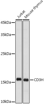
Western blot analysis of various lysates using CD3H Rabbit pAb (CAB2058) at 1:500 dilution. Secondary antibody: HRP-conjugated Goat anti-Rabbit IgG (H+L) (CABS014) at 1:10000 dilution. Lysates/proteins: 25μg per lane. Blocking buffer: 3% nonfat dry milk in TBST. Detection: ECL Enhanced Kit (AbGn00021). Exposure time: 180s.
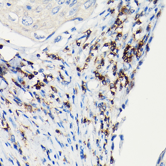
Immunohistochemistry analysis of paraffin-embedded Human esophageal cancer using CD3H Rabbit pAb (CAB2058) at dilution of 1:100 (40x lens). High pressure antigen retrieval performed with 0.01M Citrate buffer (pH 6.0) prior to IHC staining.