The CD27 Monoclonal Antibody (CAB11505) is a high-quality antibody developed for reliable detection and analysis of target proteins. CD27 is a cell surface molecule that plays a crucial role in immune regulation, particularly in activating immune responses. This antibody, generated in rabbits, is validated for use in Western blot applications and is highly reactive with human samples.CD27, also known as TNFRSF7, is a vital co-stimulatory molecule that promotes T-cell activation and proliferation. Its involvement in immune responses makes it a target of interest in immunology and cancer research.
This antibody is validated for use in WB, ELISA, IF-P applications and has demonstrated reactivity against Human, Rat samples.
Product Name:
CD27 Monoclonal Antibody
SKU:
CAB11505
Size:
20μL, 100μL
Reactivity:
Human, Rat
Clone Number:
ARC0625
Conjugate:
Unconjugated
Immunogen:
Synthetic peptide. This information is considered to be commercially sensitive.
Recommended starting concentration is 1 μg/mL. Please optimize the concentration based on your specific assay requirements.
Synonyms:
T14, S152, Tp55, TNFRSF7, S152. LPFS2, CD27
Positive Sample:
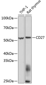
THP-1, Rat thymus
Cellular Localization:
Membrane, Single-Pass Type I Membrane Protein.
Calculated MW:
29kDa
Observed MW:
55kDa
The protein encoded by this gene is a member of the TNF-receptor superfamily. This receptor is required for generation and long-term maintenance of T cell immunity. It binds to ligand CD70, and plays a key role in regulating B-cell activation and immunoglobulin synthesis. This receptor transduces signals that lead to the activation of NF-kappaB and MAPK8/JNK. Adaptor proteins TRAF2 and TRAF5 have been shown to mediate the signaling process of this receptor. CD27-binding protein (SIVA), a proapoptotic protein, can bind to this receptor and is thought to play an important role in the apoptosis induced by this receptor.
Purification Method
Affinity purification
Gene ID
939
RRID
AB_2861587
Buffer Information
Store at -20℃. Avoid freeze / thaw cycles. Buffer: PBS containing 50% glycerol and 0.05% BSA, preserved with proclin300 or sodium azide, pH 7.3.
Western blot analysis of various lysates using CD27 Rabbit mAb (CAB11505) at 1:1000 dilution. Secondary antibody: HRP-conjugated Goat anti-Rabbit IgG (H+L) (CABS014) at 1:10000 dilution. Lysates/proteins: 25μg per lane. Blocking buffer: 3% nonfat dry milk in TBST. Detection: ECL Basic Kit (AbGn00020). Exposure time: 3s.
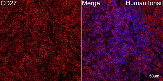
Confocal imaging of human tonsil using CD27 Rabbit mAb (CAB11505, dilution 1:100) (Red). DAPI was used for nuclear staining (blue). Objective: 40x.Perform high pressure antigen retrieval with 10 mM citrate buffer pH 6.0 before commencing with IF staining protocol.


")
")
")
")

