The CD274 Monoclonal Antibody (CAB23922) is a high-quality antibody developed for reliable detection and analysis of target proteins. This antibody, developed through monoclonal technology, exhibits high specificity and sensitivity to human CD274 protein, making it ideal for various experimental applications.CD274/PD-L1 is a critical immune checkpoint protein that plays a key role in suppressing immune responses and promoting immune tolerance. In cancer, CD274 expression on tumor cells allows them to evade the immune system's detection, making it a promising target for immunotherapy treatment strategies.
This antibody is validated for use in WB, IHC-P, IF/ICC, ELISA applications and has demonstrated reactivity against Mouse samples.
Product Name:
CD274 Monoclonal Antibody
SKU:
CAB23922
Size:
20μL, 100μL
Reactivity:
Mouse
Clone Number:
ARC61782
Conjugate:
Unconjugated
Immunogen:
Recombinant protein (or fragment).This information is considered to be commercially sensitive.
Cell Membrane, Endomembrane System, Single-Pass Type I Membrane Protein.
Calculated MW:
33kDa
Observed MW:
40-50kDa
This gene encodes an immune inhibitory receptor ligand that is expressed by hematopoietic and non-hematopoietic cells, such as T cells and B cells and various types of tumor cells. The encoded protein is a type I transmembrane protein that has immunoglobulin V-like and C-like domains. Interaction of this ligand with its receptor inhibits T-cell activation and cytokine production. During infection or inflammation of normal tissue, this interaction is important for preventing autoimmunity by maintaining homeostasis of the immune response. In tumor microenvironments, this interaction provides an immune escape for tumor cells through cytotoxic T-cell inactivation. Expression of this gene in tumor cells is considered to be prognostic in many types of human malignancies, including colon cancer and renal cell carcinoma. Alternative splicing results in multiple transcript variants.
Purification Method
Affinity purification
Gene ID
60533
Buffer Information
Store at -20℃. Avoid freeze / thaw cycles. Buffer: PBS containing 50% glycerol and 0.05% BSA, preserved with proclin300 or sodium azide, pH 7.3.
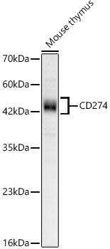
Western blot analysis of lysates from Mouse thymus, using CD274 Rabbit mAb (CAB23922) at 1:1000 dilution. Secondary antibody: HRP-conjugated Goat anti-Rabbit IgG (H+L) (CABS014) at 1:10000 dilution. Lysates/proteins: 25μg per lane. Blocking buffer: 3% nonfat dry milk in TBST. Detection: ECL Basic Kit (AbGn00020). Exposure time: 60s.
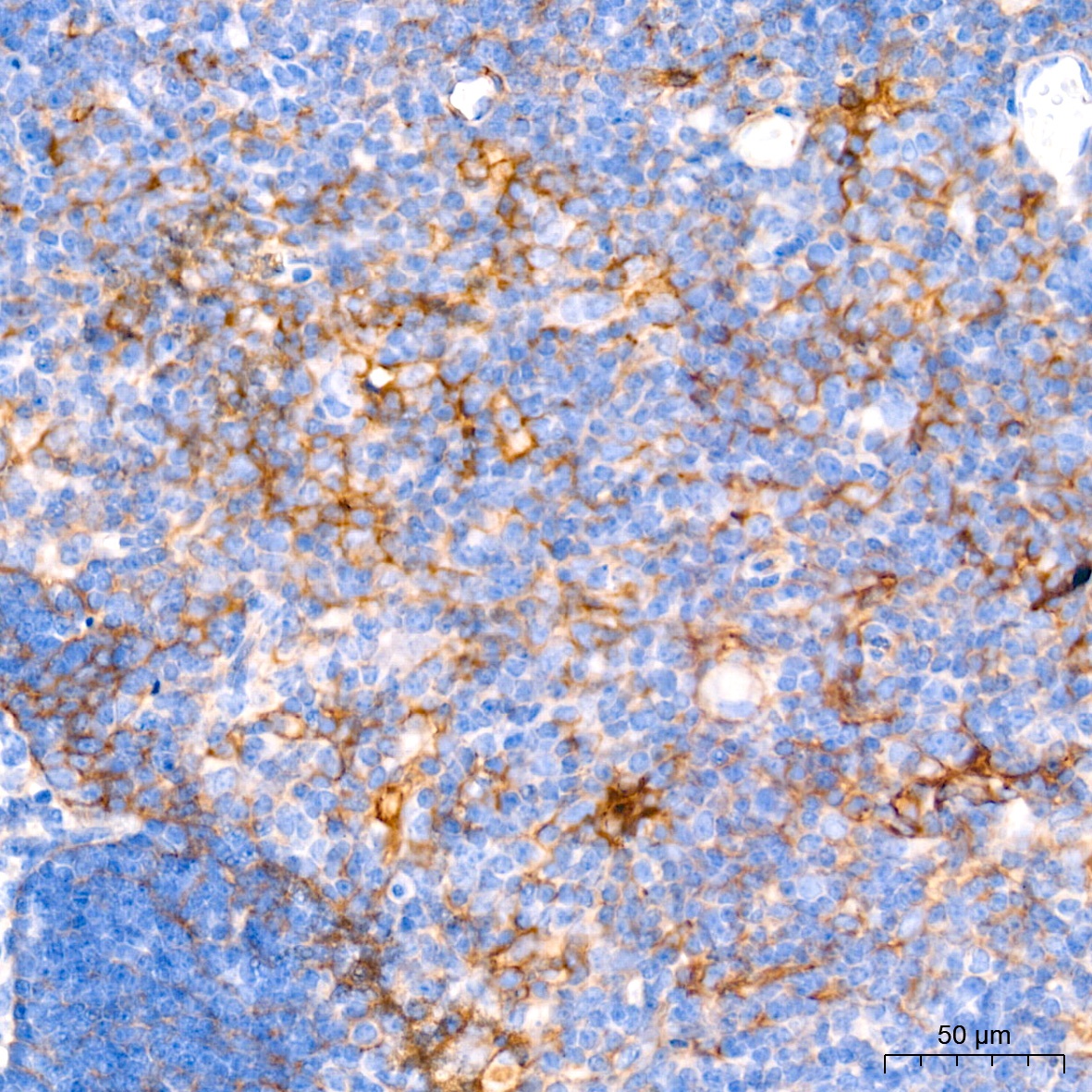
Immunohistochemistry analysis of paraffin-embedded Mouse thymus tissue using CD274 Rabbit mAb (CAB23922) at a dilution of 1:500 (40x lens). High pressure antigen retrieval performed with 0.01M Tris-EDTA Buffer (pH 9.0) prior to IHC staining.
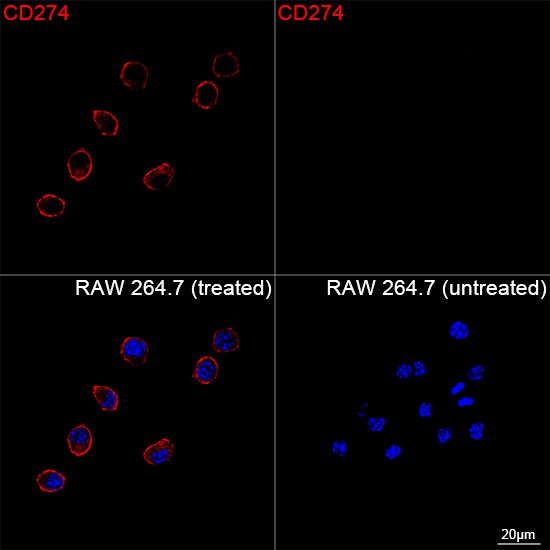
Confocal imaging of RAW 264.7 cells (treated with IFN-γ) and RAW 264.7 cells (untreated) using CD274 Rabbit mAb (CAB23922, dilution 1:200) followed by a further incubation with Cy3 Goat Anti-Rabbit IgG (H+L) (CABS007, dilution 1:500) (Red). DAPI was used for nuclear staining (Blue). Objective: 100x.
 at 1:1000 dilution. Secondary antibody: HRP Goat Anti-Rabbit IgG (H+L) at 1:10000 dilution. Lysates/proteins: 25μg per lane. Blocking buffer: 3% nonfat dry milk in TBST.")
 at 1:1000 dilution. Secondary antibody: HRP Goat Anti-Rabbit IgG (H+L) at 1:10000 dilution. Lysates/proteins: 25μg per lane. Blocking buffer: 3% nonfat dry milk in TBST.")


