The PD-L1/CD274 Antibody (CAB11273) is a high-quality antibody developed for reliable detection and analysis of target proteins. PD-L1 is known to suppress the immune response, making it a critical target for cancer immunotherapy research. This antibody, produced in rabbits, exhibits high reactivity with human samples and is validated for use in Western blot applications.By specifically binding to the CD274 protein, this antibody enables precise detection and analysis in various cell types, making it ideal for studies in cancer biology and immunology.
This antibody is validated for use in WB, IHC-P, ELISA applications and has demonstrated reactivity against Human, Mouse, Rat samples.
Product Name:
PD-L1/CD274 Antibody
SKU:
CAB11273
Size:
20μL, 100μL
Reactivity:
Human, Mouse, Rat
Conjugate:
Unconjugated
Immunogen:
Synthetic peptide. This information is considered to be commercially sensitive.
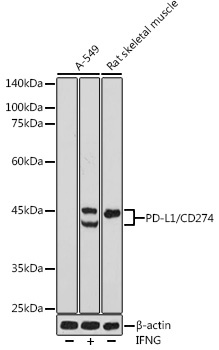
A-549 treated with IFNG, Mouse skeletal muscle, Rat skeletal muscle
Cellular Localization:
Cell Membrane, Endomembrane System, Single-Pass Type I Membrane Protein.
Calculated MW:
33kDa
Observed MW:
40-50kDa
This gene encodes an immune inhibitory receptor ligand that is expressed by hematopoietic and non-hematopoietic cells, such as T cells and B cells and various types of tumor cells. The encoded protein is a type I transmembrane protein that has immunoglobulin V-like and C-like domains. Interaction of this ligand with its receptor inhibits T-cell activation and cytokine production. During infection or inflammation of normal tissue, this interaction is important for preventing autoimmunity by maintaining homeostasis of the immune response. In tumor microenvironments, this interaction provides an immune escape for tumor cells through cytotoxic T-cell inactivation. Expression of this gene in tumor cells is considered to be prognostic in many types of human malignancies, including colon cancer and renal cell carcinoma. Alternative splicing results in multiple transcript variants.
Purification Method
Affinity purification
Gene ID
29126
RRID
AB_2758487
Buffer Information
Store at -20℃. Avoid freeze / thaw cycles. Buffer: PBS containing 50% glycerol, preserved with proclin300 or sodium azide, pH 7.3.
Western blot analysis of various lysates using PD-L1/CD274 Rabbit pAb (CAB11273) at 1:500 dilution. A-549 cells were treated with IFNG (100 ng/mL) at 37℃ for 48 hours. Secondary antibody: HRP-conjugated Goat anti-Rabbit IgG (H+L) (CABS014) at 1:10000 dilution. Lysates/proteins: 25μg per lane. Blocking buffer: 3% nonfat dry milk in TBST. Detection: ECL Basic Kit (AbGn00020). Exposure time: 180s.
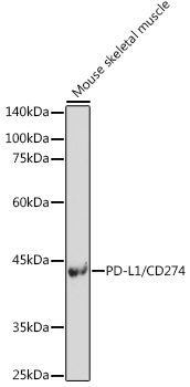
Western blot analysis of lysates from Mouse skeletal muscle, using PD-L1/CD274 Rabbit pAb (CAB11273) at 1:500 dilution. Secondary antibody: HRP-conjugated Goat anti-Rabbit IgG (H+L) (CABS014) at 1:10000 dilution. Lysates/proteins: 25μg per lane. Blocking buffer: 3% nonfat dry milk in TBST. Detection: ECL Enhanced Kit (AbGn00021). Exposure time: 180s.
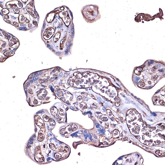
Immunohistochemistry analysis of paraffin-embedded Human placenta using PD-L1/CD274 Rabbit pAb (CAB11273) at dilution of 1:100 (40x lens). High pressure antigen retrieval performed with 0.01M Citrate buffer (pH 6.0) prior to IHC staining.


![FITC Anti-Mouse CD274/PD-L1 Antibody [10F.9G2] (AGEL2741)](https://cdn11.bigcommerce.com/s-h68l9z2lnx/images/stencil/590x590/products/229366/605234/fitc-anti-mouse-cd274pd-l1-antibody-10f.9g2-agel2741__65790.1706889767.jpg?c=2 "FITC Anti-Mouse CD274/PD-L1 Antibody [10F.9G2] (AGEL2741)")