The PD-L1/CD274 Antibody (CAB1645) is a high-quality antibody developed for reliable detection and analysis of target proteins. This antibody, generated in rabbits, is highly specific for human samples and has been validated for use in Western blot applications. By binding to CD274, researchers can detect and analyze this important immune checkpoint protein in various cell types, making it an ideal choice for studies in immunology and cancer research.CD274 is a key player in the immune system, serving as a checkpoint molecule that helps regulate immune responses and prevent excessive inflammation. Its role in immune evasion has critical implications for diseases like cancer, where tumors can exploit CD274 to evade detection and destruction by the immune system.
This antibody is validated for use in WB, IHC-P, ELISA, IF-P applications and has demonstrated reactivity against Human, Mouse, Rat samples.
Product Name:
PD-L1/CD274 Antibody
SKU:
CAB1645
Size:
20μL, 100μL
Reactivity:
Human, Mouse, Rat
Conjugate:
Unconjugated
Immunogen:
Synthetic peptide. This information is considered to be commercially sensitive.
Cell Membrane, Endomembrane System, Single-Pass Type I Membrane Protein.
Calculated MW:
33kDa
Observed MW:
40-50kDa
This gene encodes an immune inhibitory receptor ligand that is expressed by hematopoietic and non-hematopoietic cells, such as T cells and B cells and various types of tumor cells. The encoded protein is a type I transmembrane protein that has immunoglobulin V-like and C-like domains. Interaction of this ligand with its receptor inhibits T-cell activation and cytokine production. During infection or inflammation of normal tissue, this interaction is important for preventing autoimmunity by maintaining homeostasis of the immune response. In tumor microenvironments, this interaction provides an immune escape for tumor cells through cytotoxic T-cell inactivation. Expression of this gene in tumor cells is considered to be prognostic in many types of human malignancies, including colon cancer and renal cell carcinoma. Alternative splicing results in multiple transcript variants.
Purification Method
Affinity purification
Gene ID
29126
RRID
AB_2763702
Buffer Information
Store at -20℃. Avoid freeze / thaw cycles. Buffer: PBS containing 50% glycerol, preserved with proclin300 or sodium azide, pH 7.3.
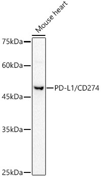
Western blot analysis of lysates from Mouse heart, using PD-L1/CD274 Rabbit pAb (CAB1645) at 1:500 dilution. Secondary antibody: HRP-conjugated Goat anti-Rabbit IgG (H+L) (CABS014) at 1:10000 dilution. Lysates/proteins: 25μg per lane. Blocking buffer: 3% nonfat dry milk in TBST. Detection: ECL Basic Kit (AbGn00020). Exposure time: 60s.
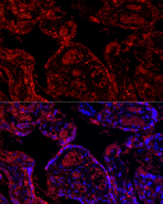
Immunofluorescence analysis of paraffin-embedded human placenta using PD-L1/CD274 Rabbit pAb (CAB1645) at dilution of 1:200 (40x lens). Secondary antibody: Cy3-conjugated Goat anti-Rabbit IgG (H+L) (CABS007) at 1:500 dilution. Blue: DAPI for nuclear staining.


![FITC Anti-Human CD274/PD-L1 Antibody [29E.2A3] (AGEL1648)](https://cdn11.bigcommerce.com/s-h68l9z2lnx/images/stencil/590x590/products/22179/607300/fitc-anti-human-cd274pd-l1-antibody-29e.2a3-agel1648__30241.1707500711.jpg?c=2 "FITC Anti-Human CD274/PD-L1 Antibody [29E.2A3] (AGEL1648)")