The CD3E Monoclonal Antibody (CAB19017) is a high-quality antibody developed for reliable detection and analysis of target proteins. This monoclonal antibody, developed in rabbits, is highly specific for human samples and has been rigorously validated for use in various applications, including Western blotting.By binding specifically to the CD3 Epsilon protein, this antibody enables accurate detection and analysis in a wide range of cell types, making it a valuable tool for researchers in the fields of immunology and cancer biology. Understanding the role of CD3 Epsilon in T cell activation and function is crucial for deciphering the mechanisms underlying immune responses and developing targeted therapies for diseases such as cancer and autoimmune disorders.
This antibody is validated for use in WB, IHC-P, IF/ICC, ELISA, IF-P, mIHC applications and has demonstrated reactivity against Human, Mouse, Rat samples.
Product Name:
CD3E Monoclonal Antibody
SKU:
CAB19017
Size:
100μL
Reactivity:
Human, Mouse, Rat
Clone Number:
ARC51750
Conjugate:
Unconjugated
Immunogen:
Synthetic peptide. This information is considered to be commercially sensitive.
Recommended starting concentration is 1 μg/mL. Please optimize the concentration based on your specific assay requirements.
Synonyms:
T3E, TCRE, IMD18, CD3epsilon, CD3E
Positive Sample:
Mouse spleen, Jurkat, MOLT-4
Cellular Localization:
Cell Membrane, Single-Pass Type I Membrane Protein.
Calculated MW:
23kDa
Observed MW:
23kDa
The protein encoded by this gene is the CD3-epsilon polypeptide, which together with CD3-gamma, -delta and -zeta, and the T-cell receptor alpha/beta and gamma/delta heterodimers, forms the T-cell receptor-CD3 complex. This complex plays an important role in coupling antigen recognition to several intracellular signal-transduction pathways. The genes encoding the epsilon, gamma and delta polypeptides are located in the same cluster on chromosome 11. The epsilon polypeptide plays an essential role in T-cell development. Defects in this gene cause immunodeficiency. This gene has also been linked to a susceptibility to type I diabetes in women.
Purification Method
Affinity purification
Gene ID
916
RRID
AB_2862509
Buffer Information
Store at -20℃. Avoid freeze / thaw cycles. Buffer: PBS containing 50% glycerol and 0.05% BSA, preserved with proclin300 or sodium azide, pH 7.3.
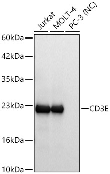
Western blot analysis of various lysates using CD3E Rabbit mAb (CAB19017) at 1:10000 dilution incubated overnight at 4℃. Secondary antibody: HRP-conjugated Goat anti-Rabbit IgG (H+L) (CABS014) at 1:10000 dilution. Lysates/proteins: 25 μg per lane. Blocking buffer: 3% nonfat dry milk in TBST. Detection: ECL Basic Kit (AbGn00020). Negative control (NC): PC-3 Exposure time: 5s.
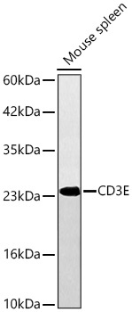
Western blot analysis of lysates from Mouse spleen using CD3E Rabbit mAb (CAB19017) at 1:10000 dilution incubated overnight at 4℃. Secondary antibody: HRP-conjugated Goat anti-Rabbit IgG (H+L) (CABS014) at 1:10000 dilution. Lysates/proteins: 25 μg per lane. Blocking buffer: 3% nonfat dry milk in TBST. Detection: ECL Basic Kit (AbGn00020). Exposure time: 10s.
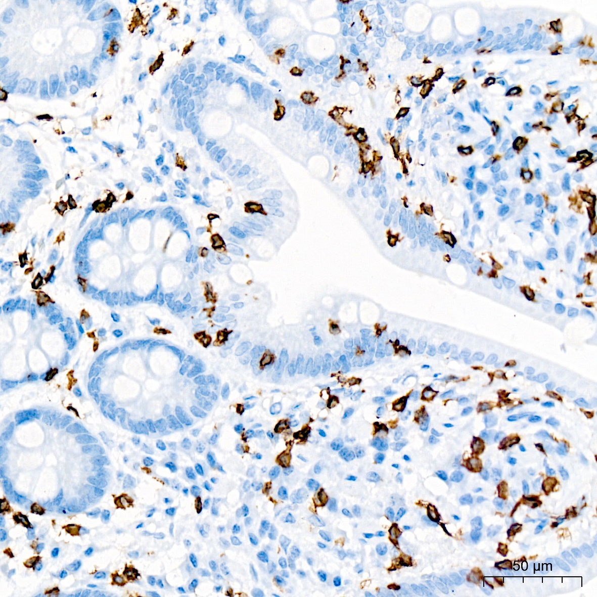
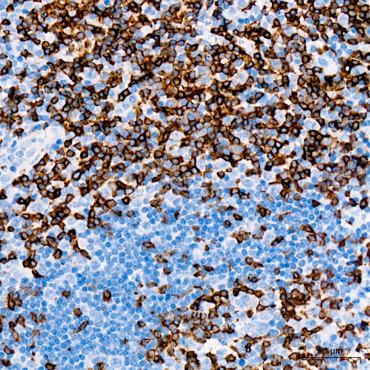
Immunohistochemistry analysis of paraffin-embedded Human small intestine tissue using CD3E Rabbit mAb (CAB19017) at a dilution of 1:2000 (40x lens). High pressure antigen retrieval was performed with 0.01 M citrate buffer (pH 6.0) prior to IHC staining.
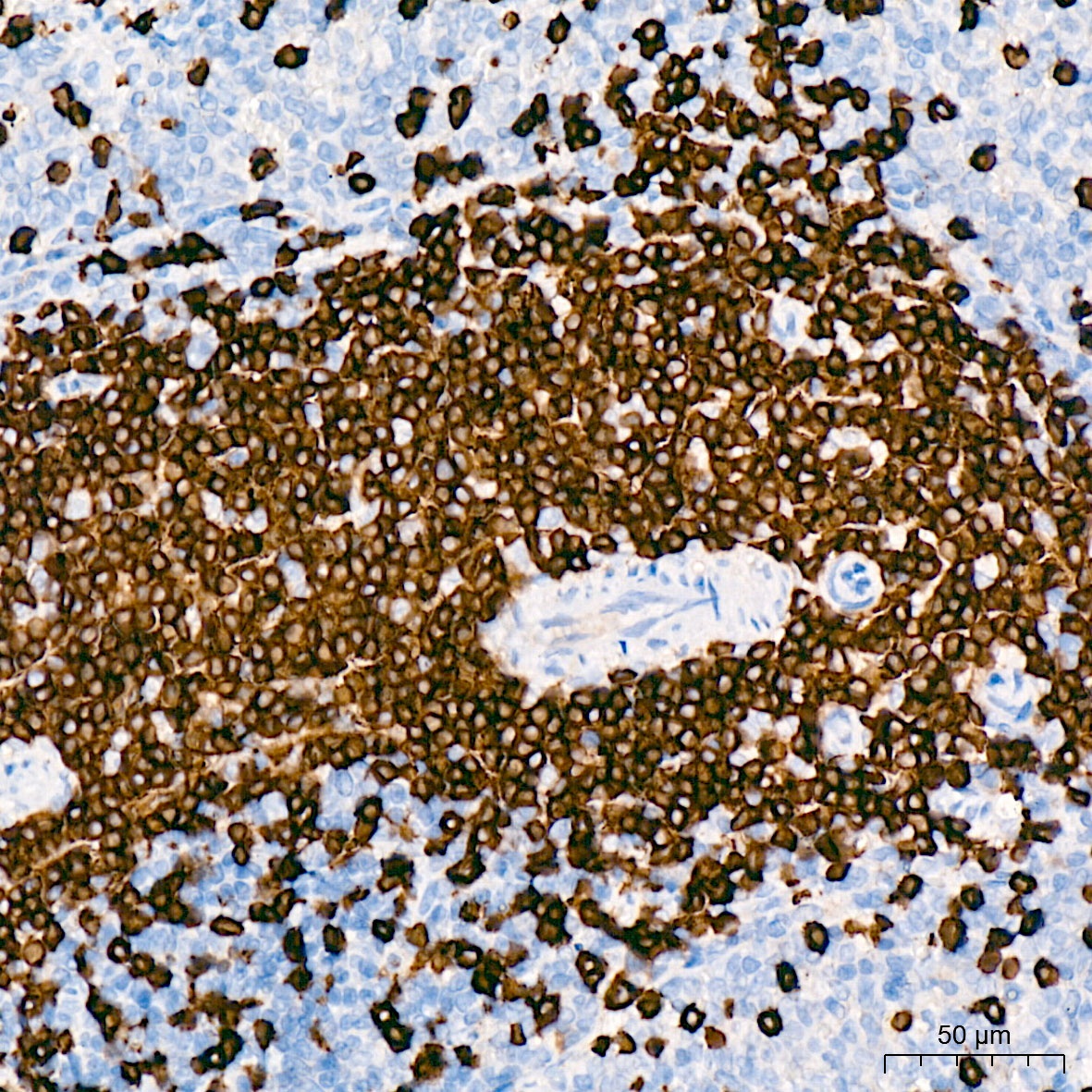
Immunohistochemistry analysis of paraffin-embedded Rat spleen tissue using CD3E Rabbit mAb (CAB19017) at a dilution of 1:2000 (40x lens). High pressure antigen retrieval was performed with 0.01 M citrate buffer (pH 6.0) prior to IHC staining.
Immunohistochemistry analysis of paraffin-embedded Human tonsil tissue using CD3E Rabbit mAb (CAB19017) at a dilution of 1:2000 (40x lens). High pressure antigen retrieval was performed with 0.01 M citrate buffer (pH 6.0) prior to IHC staining.
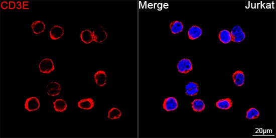
Confocal imaging of Jurkat cells using CD3E Rabbit mAb (CAB19017, dilution 1:200) followed by a further incubation with Cy3 Goat Anti-Rabbit IgG (H+L) (CABS007, dilution 1:500) (Red). DAPI was used for nuclear staining (Blue). Objective: 100x.
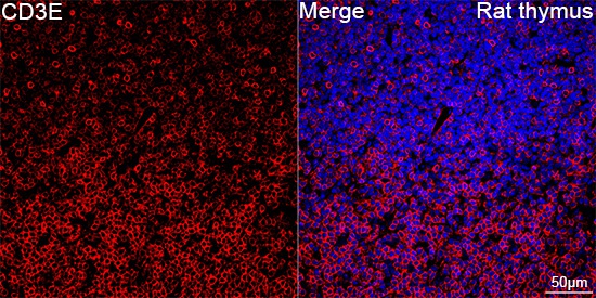
Confocal imaging of paraffin-embedded Rat thymus tissue using CD3E Rabbit mAb (CAB19017, dilution 1:200) followed by a further incubation with Cy3 Goat Anti-Rabbit IgG (H+L) (CABS007, dilution 1:500) (Red). DAPI was used for nuclear staining (Blue). High pressure antigen retrieval performed with 0.01M Citrate Buffer (pH 6.0) prior to IF staining. Objective: 40x.
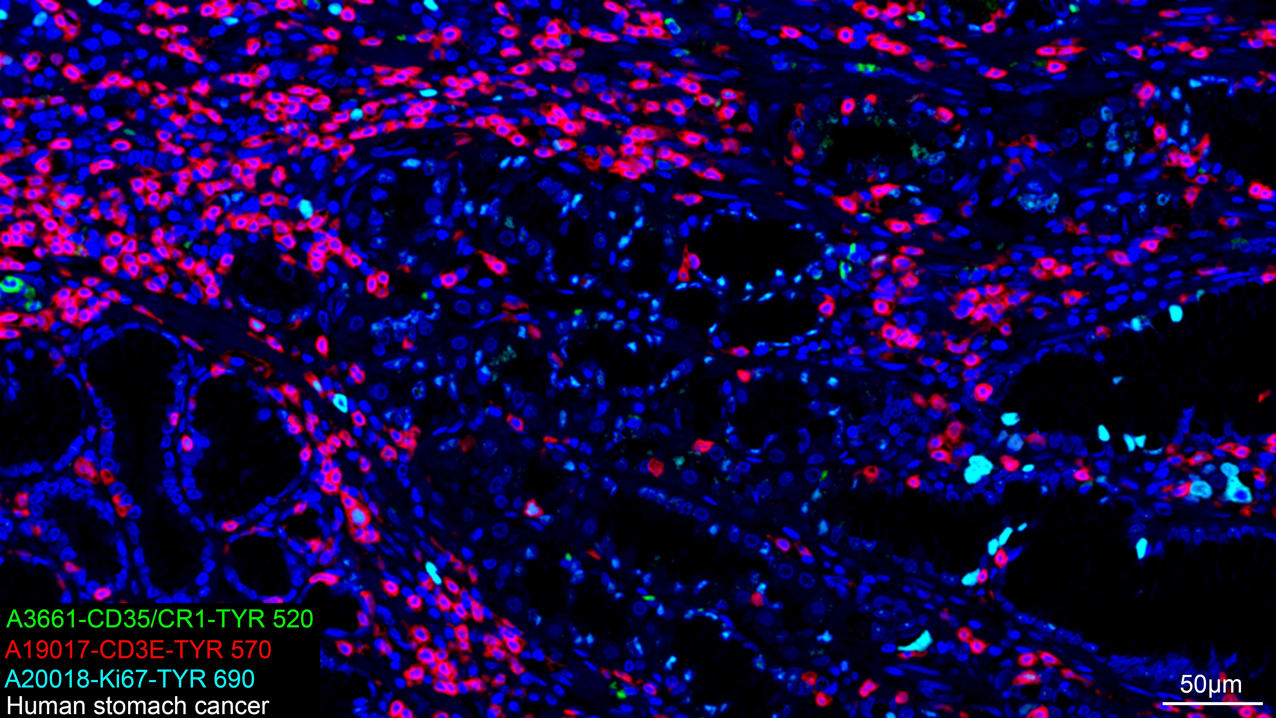
The multiplex IHC analysis on paraffin-embedded Human stomach cancer tissue using the following specific primary antibodies and tyramide signal amplification (TSA) reagents (RK05903) : CD35/CR1 Rabbit mAb (CAB3661, 1:100) with TSA-TYR-520 (Green), CD3E Rabbit mAb (CAB19017, 1:2000) with TSA-TYR-570 (Red), and Ki67 Rabbit mAb (CAB20018, 1:500) with TSA-TYR-690 (cyan). DAPI (Blue) was used for nuclear staining. Prior to multiplex IHC staining, high-pressure antigen retrieval was performed using 0.01M citrate buffer at pH 6.0. The analysis was completed using a 20x objective lens.
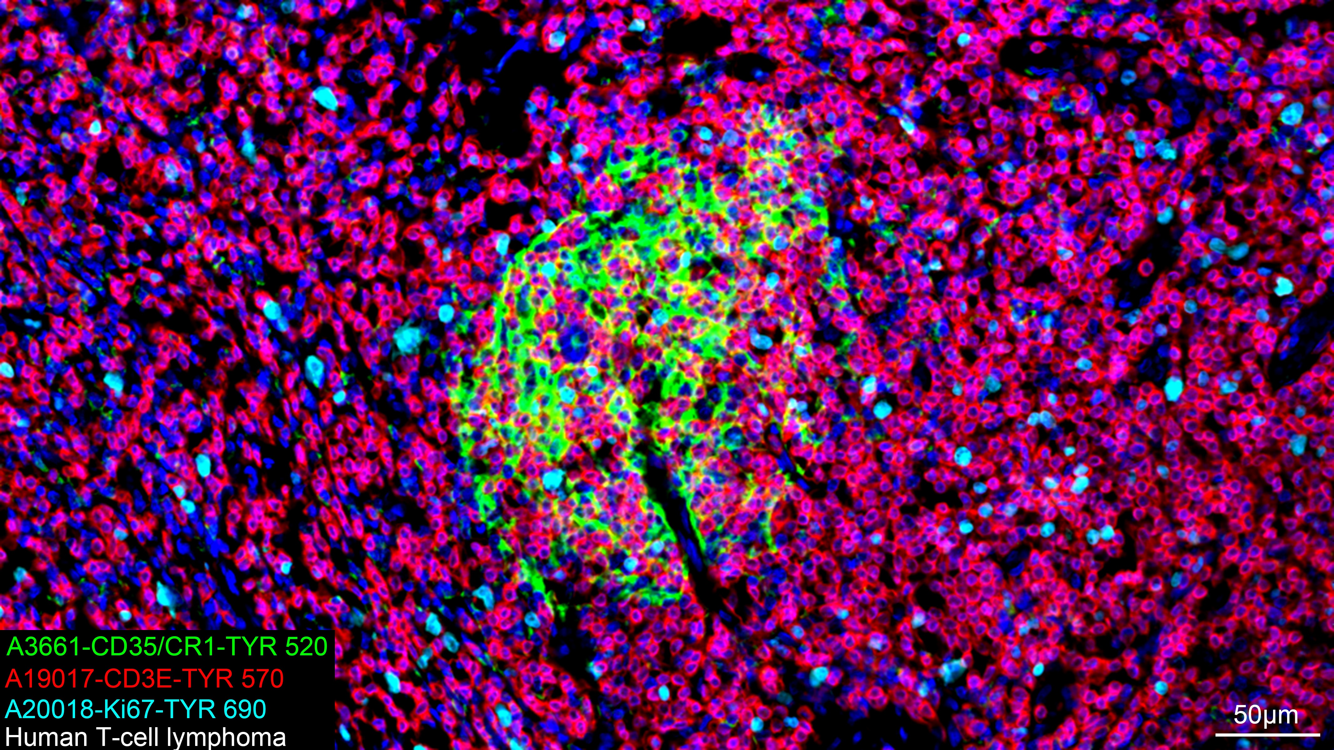
The multiplex IHC analysis on paraffin-embedded Human T-cell lymphoma tissue using the following specific primary antibodies and tyramide signal amplification (TSA) reagents (RK05903) : CD35/CR1 Rabbit mAb (CAB3661, 1:100) with TSA-TYR-520 (Green), CD3E Rabbit mAb (CAB19017, 1:2000) with TSA-TYR-570 (Red), and Ki67 Rabbit mAb (CAB20018, 1:500) with TSA-TYR-690 (cyan). DAPI (Blue) was used for nuclear staining. Prior to multiplex IHC staining, high-pressure antigen retrieval was performed using 0.01M citrate buffer at pH 6.0. The analysis was completed using a 20x objective lens.
The multiplex IHC analysis on paraffin-embedded Human T-cell lymphoma tissue using the following specific primary antibodies and tyramide signal amplification (TSA) reagents (RK05903) : CD35/CR1 Rabbit mAb (CAB3661, 1:100) with TSA-TYR-520 (Green), CD3E Rabbit mAb (CAB19017, 1:2000) with TSA-TYR-570 (Red), and Ki67 Rabbit mAb (CAB20018, 1:500) with TSA-TYR-690 (cyan). DAPI (Blue) was used for nuclear staining. Prior to multiplex IHC staining, high-pressure antigen retrieval was performed using 0.01M citrate buffer at pH 6.0. The analysis was completed using a 20x objective lens.


 (AGIM0524)")


