The CD34 Antibody (CAB0761) is a high-quality antibody developed for reliable detection and analysis of target proteins. This antibody, produced in rabbits, shows high reactivity with human samples and has been validated for use in Western blot applications. By binding specifically to the CD34 protein, this antibody allows for accurate detection and analysis in various cell types, making it ideal for research in stem cell biology, developmental biology, and cancer research.CD34 is a key marker for identifying and studying stem cells involved in hematopoiesis and vasculogenesis, making it an essential tool for researchers in the fields of regenerative medicine and tissue engineering.
This antibody is validated for use in WB, IHC-P, ELISA, IF-P applications and has demonstrated reactivity against Human, Rat samples.
Product Name:
CD34 Antibody
SKU:
CAB0761
Size:
20μL, 100μL
Reactivity:
Human, Rat
Conjugate:
Unconjugated
Immunogen:
Recombinant protein (or fragment).This information is considered to be commercially sensitive.
Recommended starting concentration is 1 μg/mL. Please optimize the concentration based on your specific assay requirements.
Synonyms:
CD34, CD34 molecule, GIG3, MORT1
Positive Sample:
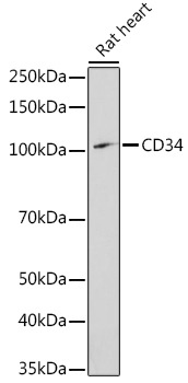
Rat heart
Cellular Localization:
Membrane, Single-Pass Type I Membrane Protein.
Calculated MW:
41kDa
Observed MW:
105kDa
The protein encoded by this gene may play a role in the attachment of stem cells to the bone marrow extracellular matrix or to stromal cells. This single-pass membrane protein is highly glycosylated and phosphorylated by protein kinase C. Two transcript variants encoding different isoforms have been found for this gene.
Purification Method
Affinity purification
Gene ID
947
RRID
AB_2757385
Buffer Information
Store at -20℃. Avoid freeze / thaw cycles. Buffer: PBS containing 50% glycerol, preserved with proclin300 or sodium azide, pH 7.3.
Western blot analysis of lysates from Rat heart, using CD34 Rabbit pAb (CAB0761) at 1:1000 dilution. Secondary antibody: HRP-conjugated Goat anti-Rabbit IgG (H+L) (CABS014) at 1:10000 dilution. Lysates/proteins: 25μg per lane. Blocking buffer: 3% nonfat dry milk in TBST. Detection: ECL Enhanced Kit (AbGn00021). Exposure time: 180s.
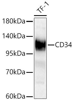
Western blot analysis of lysates from TF-1 cells using CD34 Rabbit pAb (CAB0761) at 1:1000 dilution. Secondary antibody: HRP-conjugated Goat anti-Rabbit IgG (H+L) (CABS014) at 1:10000 dilution. Lysates/proteins: 25 μg per lane. Blocking buffer: 3% nonfat dry milk in TBST. Detection: ECL Basic Kit (AbGn00020). Exposure time:30s.
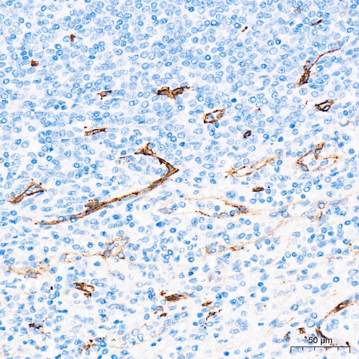
Immunohistochemistry analysis of paraffin-embedded Human spleen tissue using CD34 Rabbit pAb (CAB0761) at a dilution of 1:1000 (40x lens). High pressure antigen retrieval was performed with 0.01 M Tris-EDTA buffer (pH 9.0) prior to IHC staining.
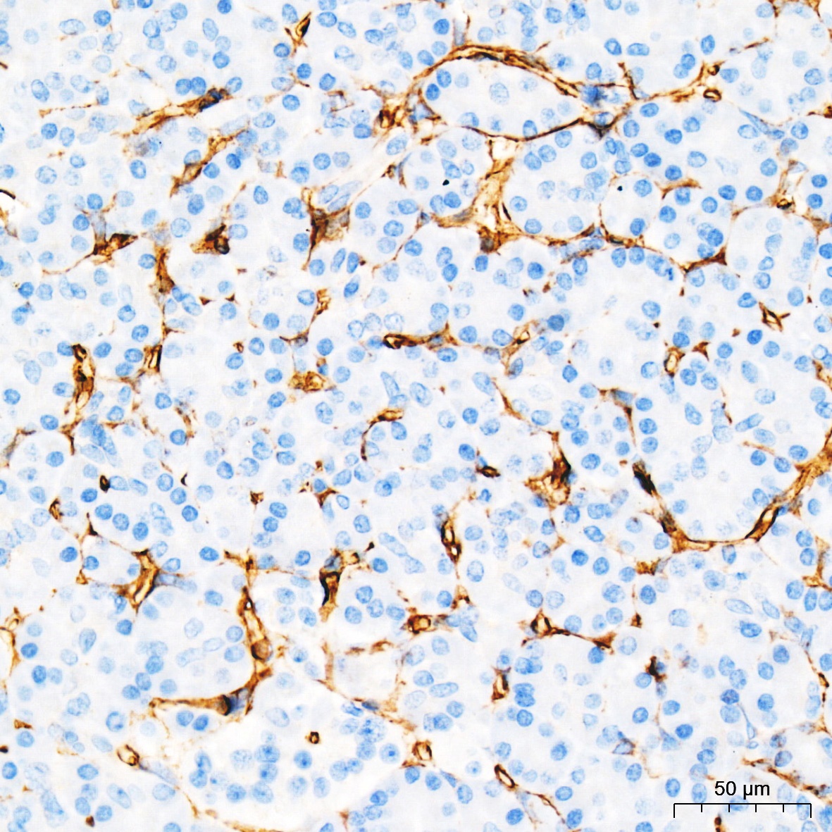
Immunohistochemistry analysis of paraffin-embedded Human pancreas tissue using CD34 Rabbit pAb (CAB0761) at a dilution of 1:1000 (40x lens). High pressure antigen retrieval was performed with 0.01 M Tris-EDTA buffer (pH 9.0) prior to IHC staining.
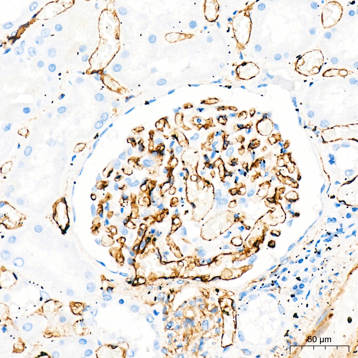
Immunohistochemistry analysis of paraffin-embedded Human kidney tissue using CD34 Rabbit pAb (CAB0761) at a dilution of 1:1000 (40x lens). High pressure antigen retrieval was performed with 0.01 M Tris-EDTA buffer (pH 9.0) prior to IHC staining.
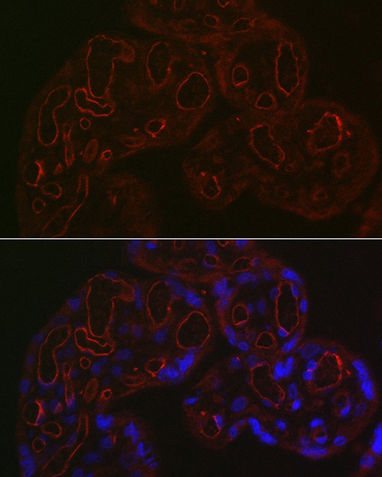
Immunofluorescence analysis of paraffin-embedded human placenta using CD34 Rabbit pAb (CAB0761) at dilution of 1:200 (40x lens). Secondary antibody: Cy3-conjugated Goat anti-Rabbit IgG (H+L) (CABS007) at 1:500 dilution. Blue: DAPI for nuclear staining.
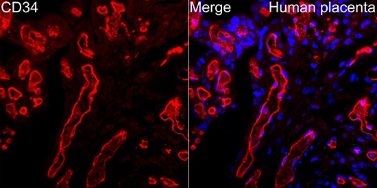
Immunofluorescence analysis of paraffin-embedded Human placenta tissue using CD34 Rabbit pAb(CAB0761) at a dilution of 1:100 (40x lens). Secondary antibody:Cy3 Goat Anti-Rabbit IgG (H+L)(CABS007) at 1:500 dilution. Blue: DAPI for nuclear staining. Perform high pressure antigen retrieval with 0.01 M citrate buffer (pH 6.0) prior to IF staining.