The CD34 Antibody (CAB13929) is a high-quality antibody developed for reliable detection and analysis of target proteins. This antibody, produced in rabbits, is highly specific and reacts with human samples, making it ideal for use in immunohistochemistry and flow cytometry applications.CD34 is a crucial marker for identifying and isolating stem cells, making this antibody essential for studies related to stem cell biology, hematopoiesis, and angiogenesis. Its ability to target CD34 protein enables the detection and analysis of CD34 expression in various tissues and cell types, providing researchers with valuable insights into cell differentiation and tissue regeneration processes.
This antibody is validated for use in WB, IHC-P, IF/ICC, ELISA applications and has demonstrated reactivity against Human, Mouse, Rat samples.
Product Name:
CD34 Antibody
SKU:
CAB13929
Size:
20μL, 100μL
Reactivity:
Human, Mouse, Rat
Conjugate:
Unconjugated
Immunogen:
Synthetic peptide. This information is considered to be commercially sensitive.
Recommended starting concentration is 1 μg/mL. Please optimize the concentration based on your specific assay requirements.
Synonyms:
CD34, CD34 molecule, GIG3, MORT1
Positive Sample:
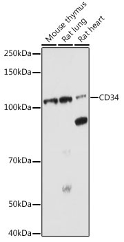
Mouse thymus, Rat lung, Rat heart
Cellular Localization:
Membrane, Single-Pass Type I Membrane Protein.
Calculated MW:
41kDa
Observed MW:
105kDa
The protein encoded by this gene may play a role in the attachment of stem cells to the bone marrow extracellular matrix or to stromal cells. This single-pass membrane protein is highly glycosylated and phosphorylated by protein kinase C. Two transcript variants encoding different isoforms have been found for this gene.
Purification Method
Affinity purification
Gene ID
947
RRID
AB_2760781
Buffer Information
Store at -20℃. Avoid freeze / thaw cycles. Buffer: PBS with 0.09% sodium azide,50% glycerol,pH7.3.
Western blot analysis of various lysates using CD34 Rabbit pAb (CAB13929) at 1:1000 dilution. Secondary antibody: HRP-conjugated Goat anti-Rabbit IgG (H+L) (CABS014) at 1:10000 dilution. Lysates/proteins: 25μg per lane. Blocking buffer: 3% nonfat dry milk in TBST. Detection: ECL Enhanced Kit (AbGn00021). Exposure time: 180s.
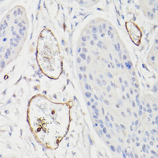
Immunohistochemistry analysis of paraffin-embedded Human esophageal cancer using CD34 Rabbit pAb (CAB13929) at dilution of 1:100 (40x lens). High pressure antigen retrieval performed with 0.01M Citrate buffer (pH 6.0) prior to IHC staining.
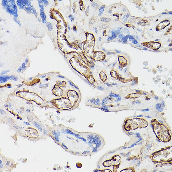
Immunohistochemistry analysis of paraffin-embedded Human placenta using CD34 Rabbit pAb (CAB13929) at dilution of 1:100 (40x lens). High pressure antigen retrieval performed with 0.01M Citrate buffer (pH 6.0) prior to IHC staining.
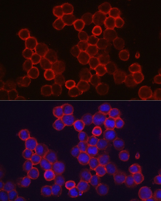
Immunofluorescence analysis of TF-1 cells using CD34 Rabbit pAb (CAB13929) at dilution of 1:100 (40x lens). Secondary antibody: Cy3-conjugated Goat anti-Rabbit IgG (H+L) (CABS007) at 1:500 dilution. Blue: DAPI for nuclear staining.