The CD3G Polyclonal Antibody (CAB1938) is a high-quality antibody developed for reliable detection and analysis of target proteins. This antibody, generated in rabbits, exhibits high reactivity with human samples and has been validated for use in Western blot applications. By binding specifically to the CD3G protein, this antibody enables accurate detection and analysis of CD3G in various cell types, making it an essential asset for studies in immunology and cancer research.
This antibody is validated for use in WB, ELISA applications and has demonstrated reactivity against Human samples.
Product Name:
CD3G Polyclonal Antibody
SKU:
CAB1938
Size:
20μL, 100μL
Reactivity:
Human
Conjugate:
Unconjugated
Immunogen:
Recombinant protein (or fragment).This information is considered to be commercially sensitive.
Recommended starting concentration is 1 μg/mL. Please optimize the concentration based on your specific assay requirements.
Synonyms:
T3G, IMD17, CD3GAMMA, CD3-GAMMA, CD3G
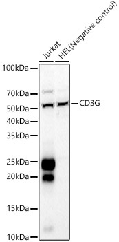
Positive Sample:
Jurkat, HEL(Negative control)
Cellular Localization:
Membrane, Single-Pass Type I Membrane Protein.
Calculated MW:
20kDa
Observed MW:
18-28kDa
The protein encoded by this gene is the CD3-gamma polypeptide, which together with CD3-epsilon, -delta and -zeta, and the T-cell receptor alpha/beta and gamma/delta heterodimers, forms the T-cell receptor-CD3 complex. This complex plays an important role in coupling antigen recognition to several intracellular signal-transduction pathways. The genes encoding the epsilon, gamma and delta polypeptides are located in the same cluster on chromosome 11. Defects in this gene are associated with T cell immunodeficiency.
Purification Method
Affinity purification
Gene ID
917
RRID
AB_2763964
Buffer Information
Store at -20℃. Avoid freeze / thaw cycles. Buffer: PBS containing 50% glycerol, preserved with proclin300 or sodium azide, pH 7.3.
Western blot analysis of various lysates, using CD3G Rabbit pAb (CAB1938) at 1:600 dilution. Secondary antibody: HRP-conjugated Goat anti-Rabbit IgG (H+L) (CABS014) at 1:10000 dilution. Lysates/proteins: 25μg per lane. Blocking buffer: 3% nonfat dry milk in TBST. Detection: ECL Basic Kit (AbGn00020). Exposure time: 45s.


")
")