The CD80 Antibody (CAB16039) is a high-quality antibody developed for reliable detection and analysis of target proteins. This antibody, produced in rabbits, exhibits high reactivity with human samples and has been validated for use in Western blot applications. By binding to the CD80 protein, this antibody enables precise detection and analysis in various cell types, making it ideal for investigations in immunology and cancer research.CD80, also known as B7-1, is a critical component of the immune system, providing essential signals for T cell activation and function. Its role in regulating immune responses makes it a valuable target for research into autoimmune diseases, transplantation, and infectious diseases.
This antibody is validated for use in WB, IHC-P, ELISA, IF-P applications and has demonstrated reactivity against Human, Mouse, Rat samples.
Product Name:
CD80 Antibody
SKU:
CAB16039
Size:
20μL, 100μL
Reactivity:
Human, Mouse, Rat
Conjugate:
Unconjugated
Immunogen:
Synthetic peptide. This information is considered to be commercially sensitive.
Recommended starting concentration is 1 μg/mL. Please optimize the concentration based on your specific assay requirements.
Synonyms:
B7, BB1, B7-1, B7.1, LAB7, CD28LG, CD28LG1, CD80
Positive Sample:
Rat lung
Cellular Localization:
Membrane, Single-Pass Type I Membrane Protein.
Calculated MW:
33kDa
Observed MW:
50-75kDa
The protein encoded by this gene is a membrane receptor that is activated by the binding of CD28 or CTLA-4. The activated protein induces T-cell proliferation and cytokine production. This protein can act as a receptor for adenovirus subgroup B and may play a role in lupus neuropathy.
Purification Method
Affinity purification
Gene ID
941
RRID
AB_2763477
Buffer Information
Store at -20℃. Avoid freeze / thaw cycles. Buffer: PBS containing 50% glycerol, preserved with proclin300 or sodium azide, pH 7.3.
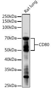
Western blot analysis of lysates from Rat lung, using CD80 Rabbit pAb (CAB16039) at 1:1000 dilution. Secondary antibody: HRP-conjugated Goat anti-Rabbit IgG (H+L) (CABS014) at 1:10000 dilution. Lysates/proteins: 25μg per lane. Blocking buffer: 3% nonfat dry milk in TBST. Detection: ECL Enhanced Kit (AbGn00021). Exposure time: 180s.
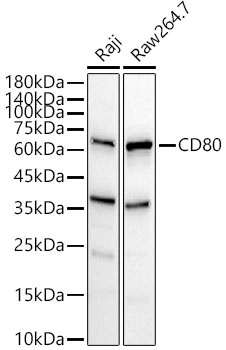
WWestern blot analysis of various lysates, using CD80 Rabbit pAb (CAB16039) at 1:900 dilution. Secondary antibody: HRP-conjugated Goat anti-Rabbit IgG (H+L) (CABS014) at 1:10000 dilution. Lysates/proteins: 25μg per lane. Blocking buffer: 3% nonfat dry milk in TBST. Detection: ECL Basic Kit (AbGn00020). Exposure time: 20s.
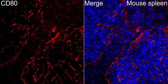
Immunofluorescence analysis of Mouse spleen tissue using CD80 Rabbit pAb (CAB16039) at a dilution of 1:200 (40x lens). Secondary antibody: Cy3-conjugated Goat anti-Rabbit IgG (H+L)(CABS007) at 1:500 dilution. Blue: DAPI for nuclear staining. High pressure antigen retrieval performed with 0.01M Citrate Buffer (pH 6.0) prior to IF staining.