The CDC40 Monoclonal Antibody (CAB19794) is a high-quality antibody developed for reliable detection and analysis of target proteins. This antibody, raised in rabbits, is highly reactive with human samples and has been validated for use in Western blot applications. It specifically binds to the CDC40 protein, enabling accurate detection and analysis in various cell types, making it an ideal tool for studies in molecular biology, cancer research, and genetic diseases.CDC40, also known as cell division cycle 40, plays a crucial role in ensuring accurate DNA replication and cell division.
This antibody is validated for use in WB, IHC-P, ELISA applications and has demonstrated reactivity against Human, Mouse, Rat samples.
Product Name:
CDC40 Monoclonal Antibody
SKU:
CAB19794
Size:
20μL, 100μL
Reactivity:
Human, Mouse, Rat
Clone Number:
ARC2321
Conjugate:
Unconjugated
Immunogen:
Synthetic peptide. This information is considered to be commercially sensitive.
Recommended starting concentration is 1 μg/mL. Please optimize the concentration based on your specific assay requirements.
Synonyms:
EHB3, PCH15, PRP17, PRPF17, CDC40
Positive Sample:
Mouse lung, HeLa, HCT 116, 293T, Rat lung
Cellular Localization:
Nuclear Speck, Nucleoplasm.
Calculated MW:
66kDa
Observed MW:
75kDa
Pre-mRNA splicing occurs in two sequential transesterification steps. The protein encoded by this gene is found to be essential for the catalytic step II in pre-mRNA splicing process. It is found in the spliceosome, and contains seven WD repeats, which function in protein-protein interactions. This protein has a sequence similarity to yeast Prp17 protein, which functions in two different cellular processes: pre-mRNA splicing and cell cycle progression. It suggests that this protein may play a role in cell cycle progression.
Purification Method
Affinity purification
Gene ID
51362
Buffer Information
Store at -20℃. Avoid freeze / thaw cycles. Buffer: PBS containing 50% glycerol and 0.05% BSA, preserved with proclin300 or sodium azide, pH 7.3.
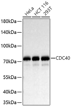
Western blot analysis of various lysates using CDC40 Rabbit mAb (CAB19794) at 1:1000 dilution incubated at room temperature for 1.5 hours. Secondary antibody: HRP-conjugated Goat anti-Rabbit IgG (H+L) (CABS014) at 1:10000 dilution. Lysates/proteins: 25 μg per lane. Blocking buffer: 3% nonfat dry milk in TBST. Detection: ECL Basic Kit (AbGn00020). Exposure time: 20 s.
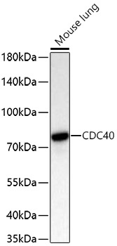
Western blot analysis of lysates from Mouse lung using CDC40 Rabbit mAb (CAB19794) at 1:1000 dilution incubated at room temperature for 1.5 hours. Secondary antibody: HRP-conjugated Goat anti-Rabbit IgG (H+L) (CABS014) at 1:10000 dilution. Lysates/proteins: 25 μg per lane. Blocking buffer: 3% nonfat dry milk in TBST. Detection: ECL Basic Kit (AbGn00020). Exposure time: 45 s.
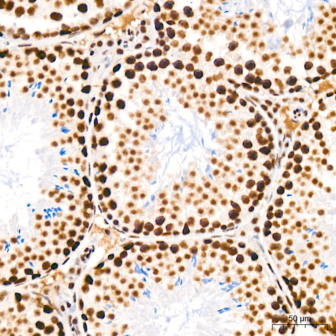
Immunohistochemistry analysis of paraffin-embedded Rat testis tissue using CDC40 Rabbit mAb (CAB19794) at a dilution of 1:200 (40x lens). High pressure antigen retrieval was performed with 0.01 M citrate buffer (pH 6.0) prior to IHC staining.
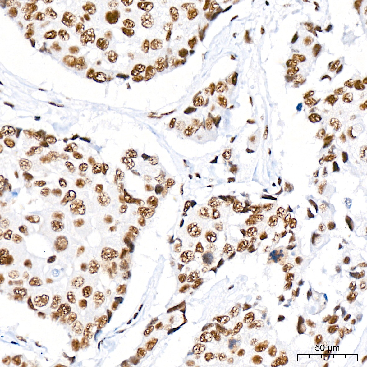
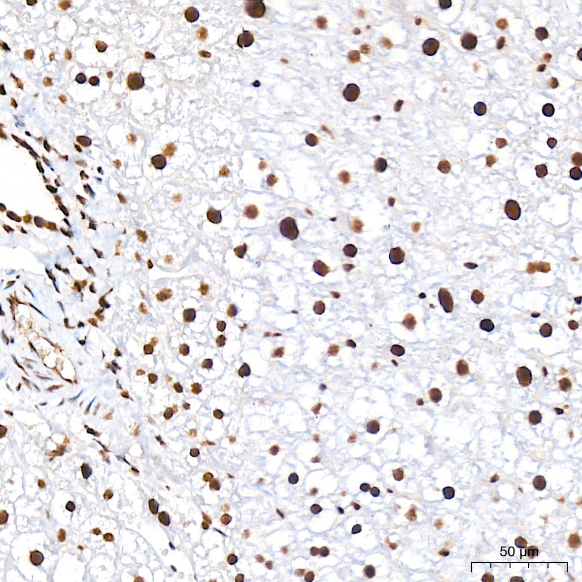
Immunohistochemistry analysis of paraffin-embedded Human breast cancer tissue using CDC40 Rabbit mAb (CAB19794) at a dilution of 1:200 (40x lens). High pressure antigen retrieval was performed with 0.01 M citrate buffer (pH 6.0) prior to IHC staining.
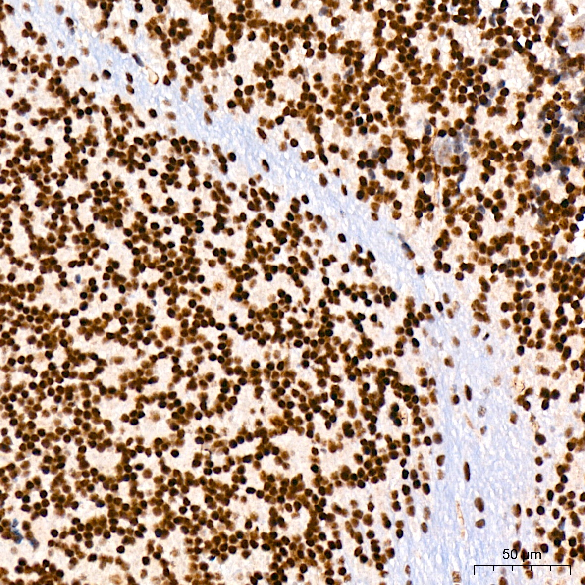
Immunohistochemistry analysis of paraffin-embedded Mouse brain tissue using CDC40 Rabbit mAb (CAB19794) at a dilution of 1:200 (40x lens). High pressure antigen retrieval was performed with 0.01 M citrate buffer (pH 6.0) prior to IHC staining.
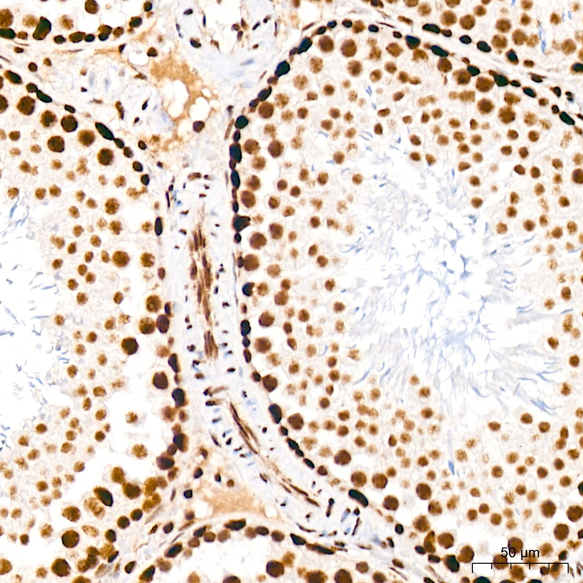
Immunohistochemistry analysis of paraffin-embedded Mouse testis tissue using CDC40 Rabbit mAb (CAB19794) at a dilution of 1:200 (40x lens). High pressure antigen retrieval was performed with 0.01 M citrate buffer (pH 6.0) prior to IHC staining.
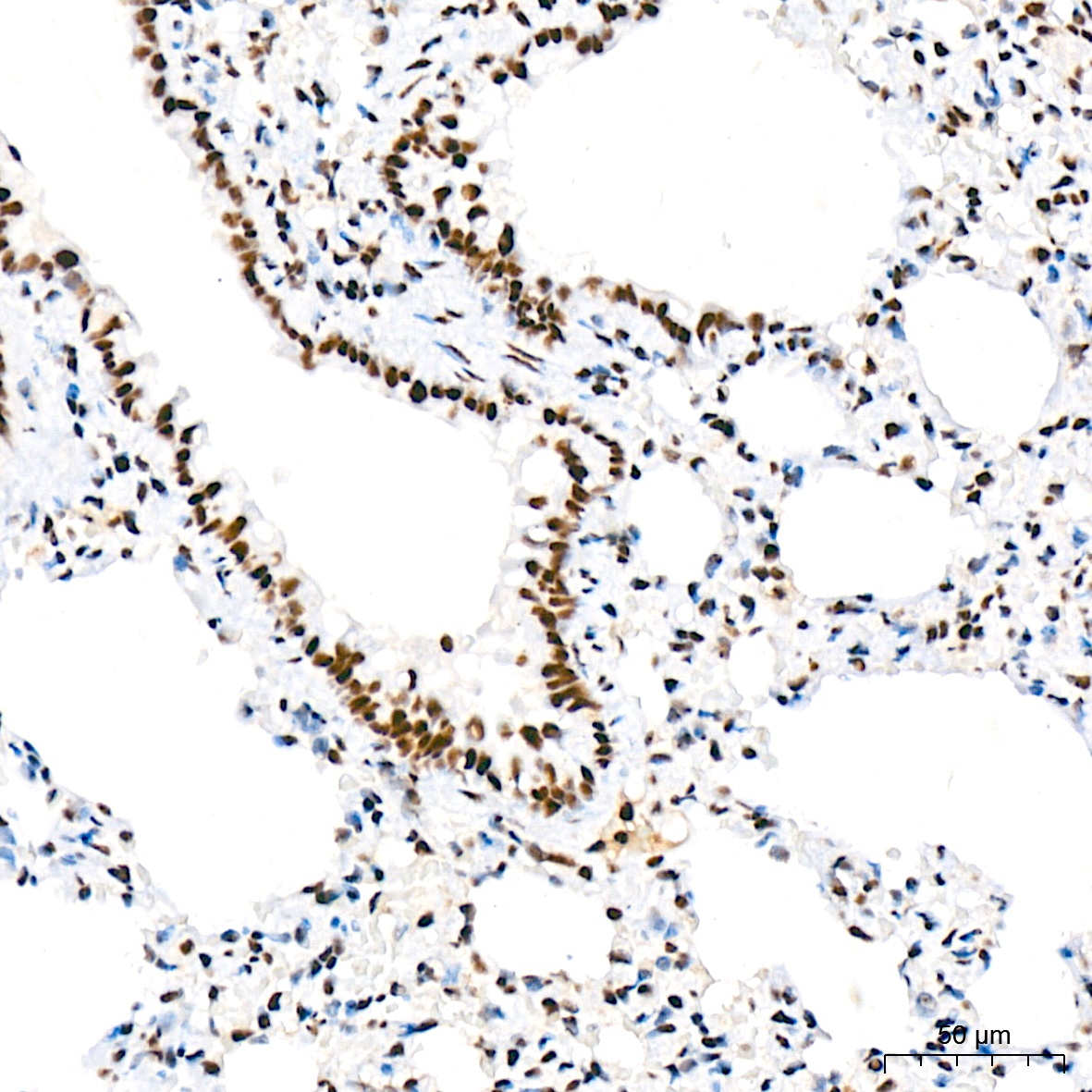
Immunohistochemistry analysis of paraffin-embedded Rat lung tissue using CDC40 Rabbit mAb (CAB19794) at a dilution of 1:200 (40x lens). High pressure antigen retrieval was performed with 0.01 M citrate buffer (pH 6.0) prior to IHC staining.
Immunohistochemistry analysis of paraffin-embedded Mouse liver tissue using CDC40 Rabbit mAb (CAB19794) at a dilution of 1:200 (40x lens). High pressure antigen retrieval was performed with 0.01 M citrate buffer (pH 6.0) prior to IHC staining.
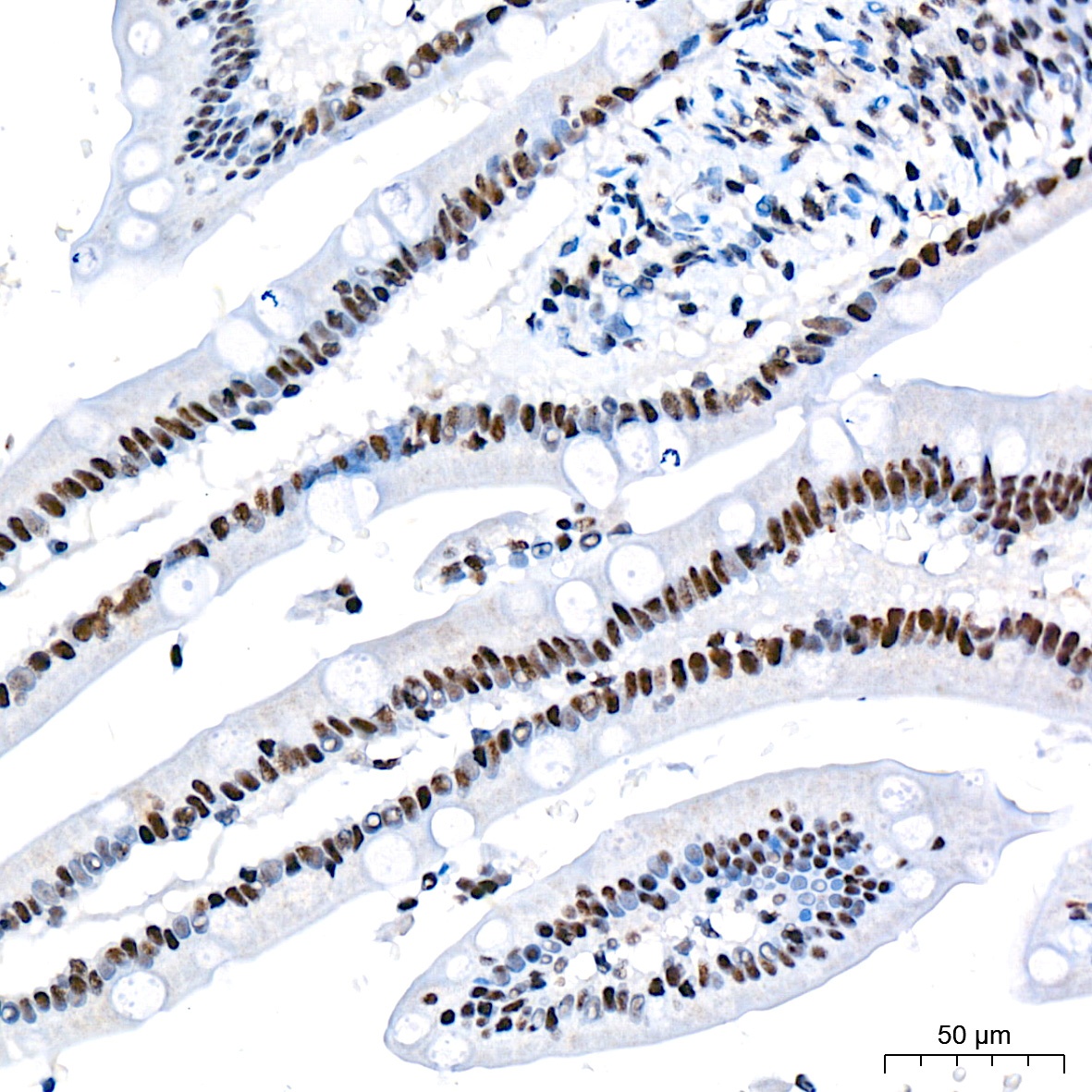
Immunohistochemistry analysis of paraffin-embedded Human small intestine tissue using CDC40 Rabbit mAb (CAB19794) at a dilution of 1:200 (40x lens). High pressure antigen retrieval was performed with 0.01 M citrate buffer (pH 6.0) prior to IHC staining.
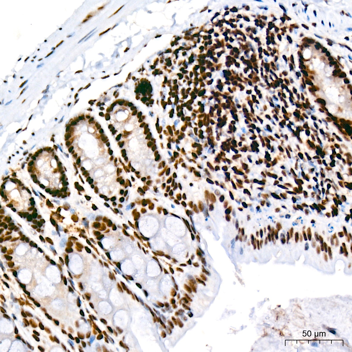
Immunohistochemistry analysis of paraffin-embedded Mouse colon tissue using CDC40 Rabbit mAb (CAB19794) at a dilution of 1:200 (40x lens). High pressure antigen retrieval was performed with 0.01 M citrate buffer (pH 6.0) prior to IHC staining.