The CENPF Antibody (CAB18644) is a high-quality antibody developed for reliable detection and analysis of target proteins. This antibody, produced in rabbits, exhibits high reactivity with human samples and has been validated for Western blot applications.CENPF is a protein that localizes to the kinetochore during mitosis and is crucial for maintaining proper chromosome alignment and segregation. Its dysregulation can lead to chromosomal instability and contribute to tumorigenesis. Therefore, understanding the function and regulation of CENPF is vital for advancing our knowledge of cell division processes and identifying potential targets for cancer therapy.
This antibody is validated for use in WB, ELISA applications and has demonstrated reactivity against Human, Mouse samples.
Product Name:
CENPF Antibody
SKU:
CAB18644
Size:
20μL, 100μL
Reactivity:
Human, Mouse
Immunogen:
Recombinant protein (or fragment).This information is considered to be commercially sensitive.
Recommended starting concentration is 1 μg/mL. Please optimize the concentration based on your specific assay requirements.
Synonyms:
CENF, hcp-1, CILD31, STROMS, PRO1779, CENPF
Positive Sample:
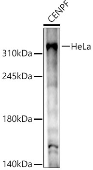
HeLa
Cellular Localization:
Axoneme, Centrosome, Ciliary Basal Body, Ciliary Transition Fiber, Cytoplasm, Cytosol, Nuclear Envelope, Nuclear Matrix, Nucleoplasm, Nucleus, Perinuclear Region Of Cytoplasm, Pronucleus, Spindle, Spindle Pole.
Calculated MW:
358kDa
Observed MW:
330kDa
This gene encodes a protein that associates with the centromere-kinetochore complex. The protein is a component of the nuclear matrix during the G2 phase of interphase. In late G2 the protein associates with the kinetochore and maintains this association through early anaphase. It localizes to the spindle midzone and the intracellular bridge in late anaphase and telophase, respectively, and is thought to be subsequently degraded. The localization of this protein suggests that it may play a role in chromosome segregation during mitotis. It is thought to form either a homodimer or heterodimer. Autoantibodies against this protein have been found in patients with cancer or graft versus host disease.
Purification Method
Affinity purification
Gene ID
1063
RRID
AB_2862382
Buffer Information
Store at -20℃. Avoid freeze / thaw cycles. Buffer: PBS containing 50% glycerol, preserved with proclin300 or sodium azide, pH 7.3.
Western blot analysis of lysates from HeLa cells, using CENPF Rabbit pAb (CAB18644) at 1:1000 dilution. Secondary antibody: HRP-conjugated Goat anti-Rabbit IgG (H+L) (CABS014) at 1:10000 dilution. Lysates/proteins: 25μg per lane. Blocking buffer: 3% nonfat dry milk in TBST. Detection: ECL Basic Kit (AbGn00020). Exposure time: 60s.