The CES1 Monoclonal Antibody (CAB11478) is a high-quality antibody developed for reliable detection and analysis of target proteins. This highly specific antibody, generated in rabbits, offers exceptional sensitivity and specificity in detecting CES1 protein levels in various cell types and tissues.CES1 is a critical enzyme involved in the metabolism of numerous drugs, including anti-cancer agents, analgesics, and antiviral medications. Understanding the expression and activity of CES1 is vital for predicting drug responses, optimizing dosages, and minimizing adverse effects in patients undergoing pharmacotherapy.
This antibody is validated for use in WB, ELISA, IF-P applications and has demonstrated reactivity against Human, Mouse, Rat samples.
Product Name:
CES1 Monoclonal Antibody
SKU:
CAB11478
Size:
20μL, 100μL
Reactivity:
Human, Mouse, Rat
Clone Number:
ARC0613
Conjugate:
Unconjugated
Immunogen:
Synthetic peptide. This information is considered to be commercially sensitive.
Hep G2, Mouse liver, Mouse kidney, Rat lung, Rat liver, Rat kidney
Cellular Localization:
Endoplasmic Reticulum Lumen.
Calculated MW:
63kDa
Observed MW:
63kDa
This gene encodes a member of the carboxylesterase large family. The family members are responsible for the hydrolysis or transesterification of various xenobiotics, such as cocaine and heroin, and endogenous substrates with ester, thioester, or amide bonds. They may participate in fatty acyl and cholesterol ester metabolism, and may play a role in the blood-brain barrier system. This enzyme is the major liver enzyme and functions in liver drug clearance. Mutations of this gene cause carboxylesterase 1 deficiency. Three transcript variants encoding three different isoforms have been found for this gene.
Purification Method
Affinity purification
Gene ID
1066
RRID
AB_2861576
Buffer Information
Store at -20℃. Avoid freeze / thaw cycles. Buffer: PBS containing 50% glycerol and 0.05% BSA, preserved with proclin300 or sodium azide, pH 7.3.
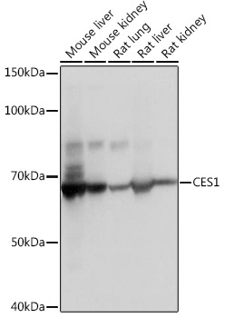
Western blot analysis of various lysates using CES1 Rabbit mAb (CAB11478) at 1:1000 dilution. Secondary antibody: HRP-conjugated Goat anti-Rabbit IgG (H+L) (CABS014) at 1:10000 dilution. Lysates/proteins: 25μg per lane. Blocking buffer: 3% nonfat dry milk in TBST. Detection: ECL Basic Kit (AbGn00020). Exposure time: 1s.
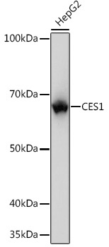
Western blot analysis of lysates from HepG2 cells, using CES1 Rabbit mAb (CAB11478) at 1:1000 dilution. Secondary antibody: HRP-conjugated Goat anti-Rabbit IgG (H+L) (CABS014) at 1:10000 dilution. Lysates/proteins: 25μg per lane. Blocking buffer: 3% nonfat dry milk in TBST. Detection: ECL Basic Kit (AbGn00020). Exposure time: 1s.
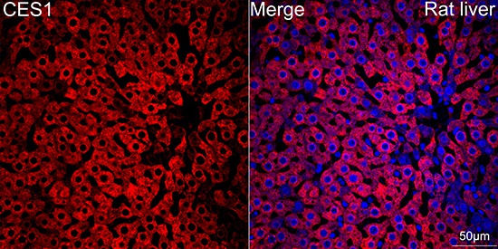
Confocal imaging of paraffin-embedded Rat liver tissue using CES1 Rabbit mAb (CAB11478, dilution 1:100) followed by a further incubation with Cy3 Goat Anti-Rabbit IgG (H+L) (CABS007, dilution 1:500) (Red). DAPI was used for nuclear staining (Blue). Objective: 40x. Perform high pressure antigen retrieval with 0.01 M citrate buffer (pH 6.0) prior to IF staining.