The [KO Validated] CHCHD2 Antibody (CAB16645) is a high-quality antibody developed for reliable detection and analysis of target proteins. This antibody, produced in rabbits, exhibits high reactivity with human samples and has been validated for use in Western blot applications. By binding specifically to the CHCHD2 protein, this antibody enables accurate detection and analysis in various cell types, making it ideal for investigations in the fields of mitochondrial biology and neurodegenerative diseases.CHCHD2 is known to play a crucial role in maintaining mitochondrial integrity and function, making it a key target for research into conditions such as neurodegenerative disorders, metabolic diseases, and aging.
This antibody is validated for use in WB, IHC-P, IF/ICC, ELISA applications and has demonstrated reactivity against Human, Mouse, Rat samples.
Product Name:
[KO Validated] CHCHD2 Antibody
SKU:
CAB16645
Size:
20μL, 100μL
Reactivity:
Human, Mouse, Rat
Conjugate:
Unconjugated
Immunogen:
Recombinant protein (or fragment).This information is considered to be commercially sensitive.
Recommended starting concentration is 1 μg/mL. Please optimize the concentration based on your specific assay requirements.
Synonyms:
MNRR1, NS2TP, MIX17B, PARK22, C7orf17, D2
Positive Sample:
HT-29, 293T, HepG2, U-87MG, mouse brain
Cellular Localization:
Nucleus.
Calculated MW:
16kDa
Observed MW:
16kDa
The protein encoded by this gene belongs to a class of eukaryotic CX(9)C proteins characterized by four cysteine residues spaced ten amino acids apart from one another. These residues form disulfide linkages that define a CHCH fold. In response to stress, the protein translocates from the mitochondrial intermembrane space to the nucleus where it binds to a highly conserved 13 nucleotide oxygen responsive element in the promoter of cytochrome oxidase 4I2, a subunit of the terminal enzyme of the electron transport chain. In concert with recombination signal sequence-binding protein J, binding of this protein activates the oxygen responsive element at four percent oxygen. In addition, it has been shown that this protein is a negative regulator of mitochondria-mediated apoptosis. In response to apoptotic stimuli, mitochondrial levels of this protein decrease, allowing BCL2-associated X protein to oligomerize and activate the caspase cascade. Pseudogenes of this gene are found on multiple chromosomes. Alternative splicing results in multiple transcript variants.
Purification Method
Affinity purification
Gene ID
51142
RRID
AB_2768881
Buffer Information
Store at -20℃. Avoid freeze / thaw cycles. Buffer: PBS with 0.01% thimerosal,50% glycerol,pH7.3.
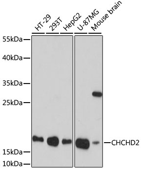
Western blot analysis of various lysates using [KO Validated] CHCHD2 Rabbit pAb (CAB16645) at 1:1000 dilution. Secondary antibody: HRP-conjugated Goat anti-Rabbit IgG (H+L) (CABS014) at 1:10000 dilution. Lysates/proteins: 25μg per lane. Blocking buffer: 3% nonfat dry milk in TBST. Detection: ECL Basic Kit (AbGn00020). Exposure time: 90s.
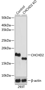
Western blot analysis of lysates from wild type (WT) and CHCHD2 knockout (KO) 293T cells, using [KO Validated] CHCHD2 Rabbit pAb (CAB16645) at 1:1000 dilution. Secondary antibody: HRP-conjugated Goat anti-Rabbit IgG (H+L) (CABS014) at 1:10000 dilution. Lysates/proteins: 25μg per lane. Blocking buffer: 3% nonfat dry milk in TBST. Detection: ECL Basic Kit (AbGn00020). Exposure time: 1s.
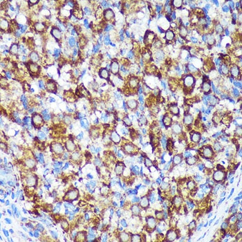
Immunohistochemistry analysis of paraffin-embedded Rat ovary using [KO Validated] CHCHD2 Rabbit pAb (CAB16645) at dilution of 1:100 (40x lens). Microwave antigen retrieval performed with 0.01M PBS Buffer (pH 7.2) prior to IHC staining.
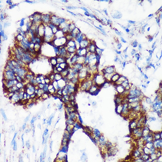
Immunohistochemistry analysis of paraffin-embedded Human colon carcinoma using [KO Validated] CHCHD2 Rabbit pAb (CAB16645) at dilution of 1:100 (40x lens). Microwave antigen retrieval performed with 0.01M PBS Buffer (pH 7.2) prior to IHC staining.
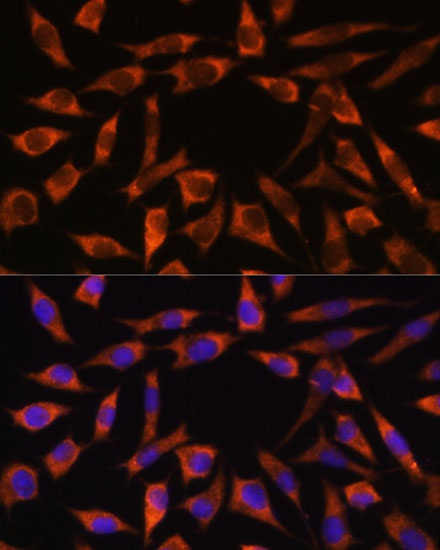
Immunofluorescence analysis of L929 cells using [KO Validated] CHCHD2 Rabbit pAb (CAB16645) at dilution of 1:100. Secondary antibody: Cy3-conjugated Goat anti-Rabbit IgG (H+L) (CABS007) at 1:500 dilution. Blue: DAPI for nuclear staining.
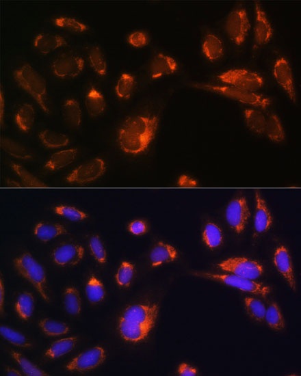
Immunofluorescence analysis of U-2 OS cells using [KO Validated] CHCHD2 Rabbit pAb (CAB16645) at dilution of 1:100. Secondary antibody: Cy3-conjugated Goat anti-Rabbit IgG (H+L) (CABS007) at 1:500 dilution. Blue: DAPI for nuclear staining.