The YKL-40/CHI3L1 Antibody (CAB3166) is a high-quality antibody developed for reliable detection and analysis of target proteins. This antibody, generated in rabbits, exhibits high reactivity with human samples and has been validated for use in Western blot applications. By binding specifically to CHI3L1, this antibody enables the detection and analysis of the protein in various cell types, making it an ideal choice for studies in immunology and cancer research.CHI3L1, also known as YKL-40, is a chitinase-like protein that is upregulated in response to inflammation and tissue injury.
This antibody is validated for use in WB, IHC-P, IF/ICC, ELISA applications and has demonstrated reactivity against Human, Mouse, Rat samples.
Product Name:
YKL-40/CHI3L1 Antibody
SKU:
CAB3166
Size:
20μL, 100μL
Reactivity:
Human, Mouse, Rat
Conjugate:
Unconjugated
Immunogen:
Recombinant protein (or fragment).This information is considered to be commercially sensitive.
Chitinases catalyze the hydrolysis of chitin, which is an abundant glycopolymer found in insect exoskeletons and fungal cell walls. The glycoside hydrolase 18 family of chitinases includes eight human family members. This gene encodes a glycoprotein member of the glycosyl hydrolase 18 family. The protein lacks chitinase activity and is secreted by activated macrophages, chondrocytes, neutrophils and synovial cells. The protein is thought to play a role in the process of inflammation and tissue remodeling.
Purification Method
Affinity purification
Gene ID
1116
RRID
AB_2764957
Buffer Information
Store at -20℃. Avoid freeze / thaw cycles. Buffer: PBS containing 50% glycerol, preserved with proclin300 or sodium azide, pH 7.3.
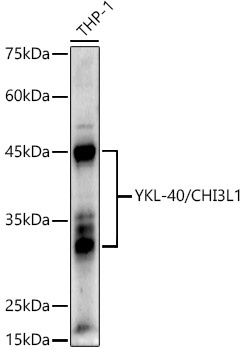
Western blot analysis of lysates from THP-1 cells, using YKL-40/CHI3L1 Rabbit pAb (CAB3166) at 1:1000 dilution. Secondary antibody: HRP-conjugated Goat anti-Rabbit IgG (H+L) (CABS014) at 1:10000 dilution. Lysates/proteins: 25μg per lane. Blocking buffer: 3% nonfat dry milk in TBST. Detection: ECL Enhanced Kit (AbGn00021). Exposure time: 180s.
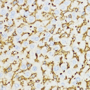
Immunohistochemistry analysis of paraffin-embedded Mouse liver using YKL-40/CHI3L1 Rabbit pAb (CAB3166) at dilution of 1:100 (40x lens). Microwave antigen retrieval performed with 0.01M PBS Buffer (pH 7.2) prior to IHC staining.
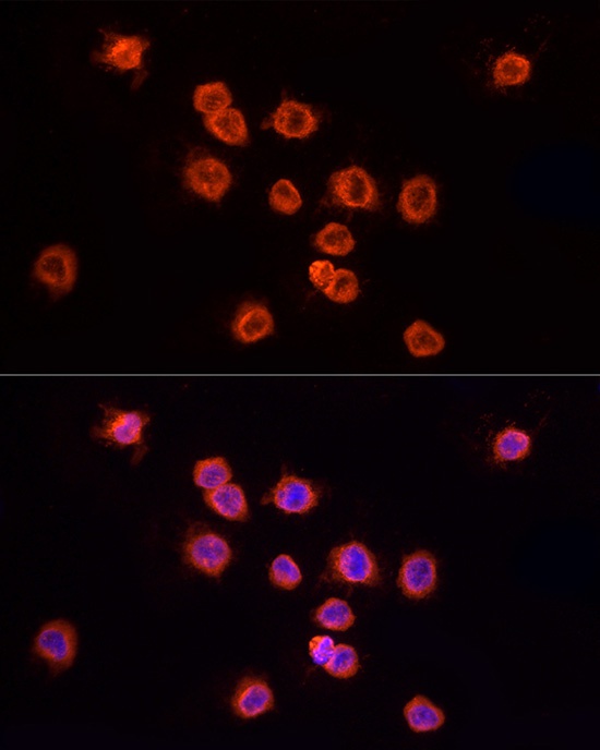
Immunofluorescence analysis of THP-1 cells using YKL-40/CHI3L1 Rabbit pAb (CAB3166) at dilution of 1:100 (40x lens). Secondary antibody: Cy3-conjugated Goat anti-Rabbit IgG (H+L) (CABS007) at 1:500 dilution. Blue: DAPI for nuclear staining.


")
")

