The CISD1 Antibody (CAB10317) is a high-quality antibody developed for reliable detection and analysis of target proteins. This antibody, produced in rabbits, is specifically designed to target CISD1 in human samples and has been validated for use in Western blot applications. By binding to CISD1, this antibody allows for the detection and analysis of the protein in various cell types, making it ideal for studies in mitochondrial biology and iron metabolism.CISD1, also known as CDGSH iron sulfur domain 1, plays a crucial role in maintaining cellular iron levels and protecting cells from oxidative damage.
This antibody is validated for use in WB, IF/ICC, ELISA applications and has demonstrated reactivity against Mouse, Rat samples.
Product Name:
CISD1 Antibody
SKU:
CAB10317
Size:
20μL, 100μL
Reactivity:
Mouse, Rat
Conjugate:
Unconjugated
Immunogen:
Recombinant protein (or fragment).This information is considered to be commercially sensitive.
Recommended starting concentration is 1 μg/mL. Please optimize the concentration based on your specific assay requirements.
Synonyms:
ZCD1, MDS029, C10orf70, mitoNEET, CISD1
Positive Sample:
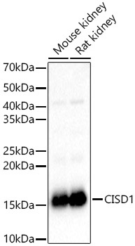
Mouse kidney, Rat kidney
Cellular Localization:
Mitochondrion Outer Membrane, Single-Pass Type Iii Membrane Protein.
Calculated MW:
12kDa
Observed MW:
16kDa
This gene encodes a protein with a CDGSH iron-sulfur domain and has been shown to bind a redox-active [2Fe-2S] cluster. The encoded protein has been localized to the outer membrane of mitochondria and is thought to play a role in regulation of oxidation. Genes encoding similar proteins are located on chromosomes 4 and 17, and a pseudogene of this gene is located on chromosome 2.
Purification Method
Affinity purification
Gene ID
55847
RRID
AB_2757861
Buffer Information
Store at -20℃. Avoid freeze / thaw cycles. Buffer: PBS containing 50% glycerol, preserved with proclin300 or sodium azide, pH 7.3.
Western blot analysis of various lysates, using CISD1 Rabbit pAb (CAB10317) at 1:1000 dilution. Secondary antibody: HRP-conjugated Goat anti-Rabbit IgG (H+L) (CABS014) at 1:10000 dilution. Lysates/proteins: 25μg per lane. Blocking buffer: 3% nonfat dry milk in TBST. Detection: ECL Basic Kit (AbGn00020). Exposure time: 1s.
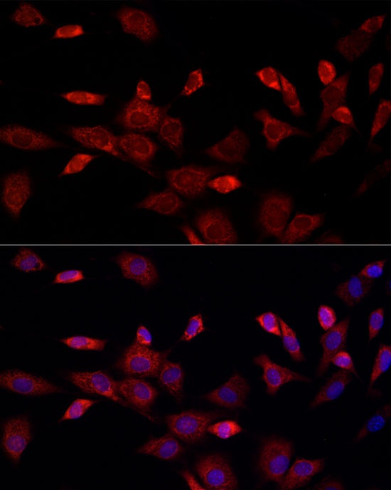
Immunofluorescence analysis of NIH/3T3 cells using CISD1 Rabbit pAb (CAB10317) at dilution of 1:100 (40x lens). Secondary antibody: Cy3-conjugated Goat anti-Rabbit IgG (H+L) (CABS007) at 1:500 dilution. Blue: DAPI for nuclear staining.