Integrin alpha V (ITGAV/CD51) Monoclonal Antibody (CAB19071)
The Integrin alpha V (ITGAV/CD51) Monoclonal Antibody (CAB19071) is a high-quality antibody developed for reliable detection and analysis of target proteins. This antibody, raised in rabbits, specifically targets the integrin alpha V protein, enabling precise detection and analysis in a variety of cell types.Integrin alpha V is known to play a critical role in processes such as cell migration, proliferation, and survival, making it a key target for studies in cell biology and cancer research. This antibody is validated for use in Western blot applications, providing researchers with a reliable tool for investigating the function and regulation of integrin alpha V in various biological contexts.
This antibody is validated for use in WB, IHC-P, ELISA, FC (intra) applications and has demonstrated reactivity against Human, Mouse, Rat samples.
Product Name:
Integrin alpha V (ITGAV/CD51) Monoclonal Antibody
SKU:
CAB19071
Size:
20μL, 100μL
Reactivity:
Human, Mouse, Rat
Clone Number:
ARC50621
Conjugate:
Unconjugated
Immunogen:
Synthetic peptide. This information is considered to be commercially sensitive.
Recommended starting concentration is 1 μg/mL. Please optimize the concentration based on your specific assay requirements.
Synonyms:
CD51, MSK8, VNRA, VTNR, Integrin alpha V (ITGAV/CD51)
Positive Sample:
A549, MCF7, NIH/3T3, C6, Mouse brain, Mouse kidney, Rat kidney, Rat lung
Cellular Localization:
Membrane, Single-Pass Type I Membrane Protein.
Calculated MW:
116kDa
Observed MW:
140kDa
The product of this gene belongs to the integrin alpha chain family. Integrins are heterodimeric integral membrane proteins composed of an alpha subunit and a beta subunit that function in cell surface adhesion and signaling. The encoded preproprotein is proteolytically processed to generate light and heavy chains that comprise the alpha V subunit. This subunit associates with beta 1, beta 3, beta 5, beta 6 and beta 8 subunits. The heterodimer consisting of alpha V and beta 3 subunits is also known as the vitronectin receptor. This integrin may regulate angiogenesis and cancer progression. Alternative splicing results in multiple transcript variants. Note that the integrin alpha 5 and integrin alpha V subunits are encoded by distinct genes.
Purification Method
Affinity purification
Gene ID
3685
RRID
AB_2862563
Buffer Information
Store at -20℃. Avoid freeze / thaw cycles. Buffer: PBS containing 50% glycerol and 0.05% BSA, preserved with proclin300 or sodium azide, pH 7.3.
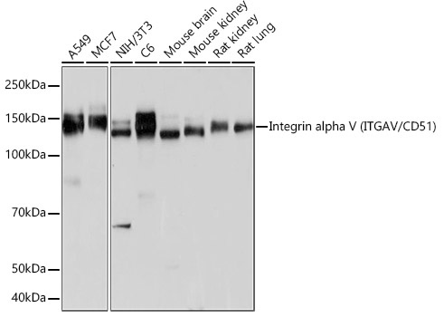
Western blot analysis of various lysates using Integrin alpha V (ITGAV/CD51) Rabbit mAb (CAB19071) at 1:1000 dilution. Secondary antibody: HRP-conjugated Goat anti-Rabbit IgG (H+L) (CABS014) at 1:10000 dilution. Lysates/proteins: 25μg per lane. Blocking buffer: 3% nonfat dry milk in TBST. Detection: ECL Basic Kit (AbGn00020). Exposure time: 1s.
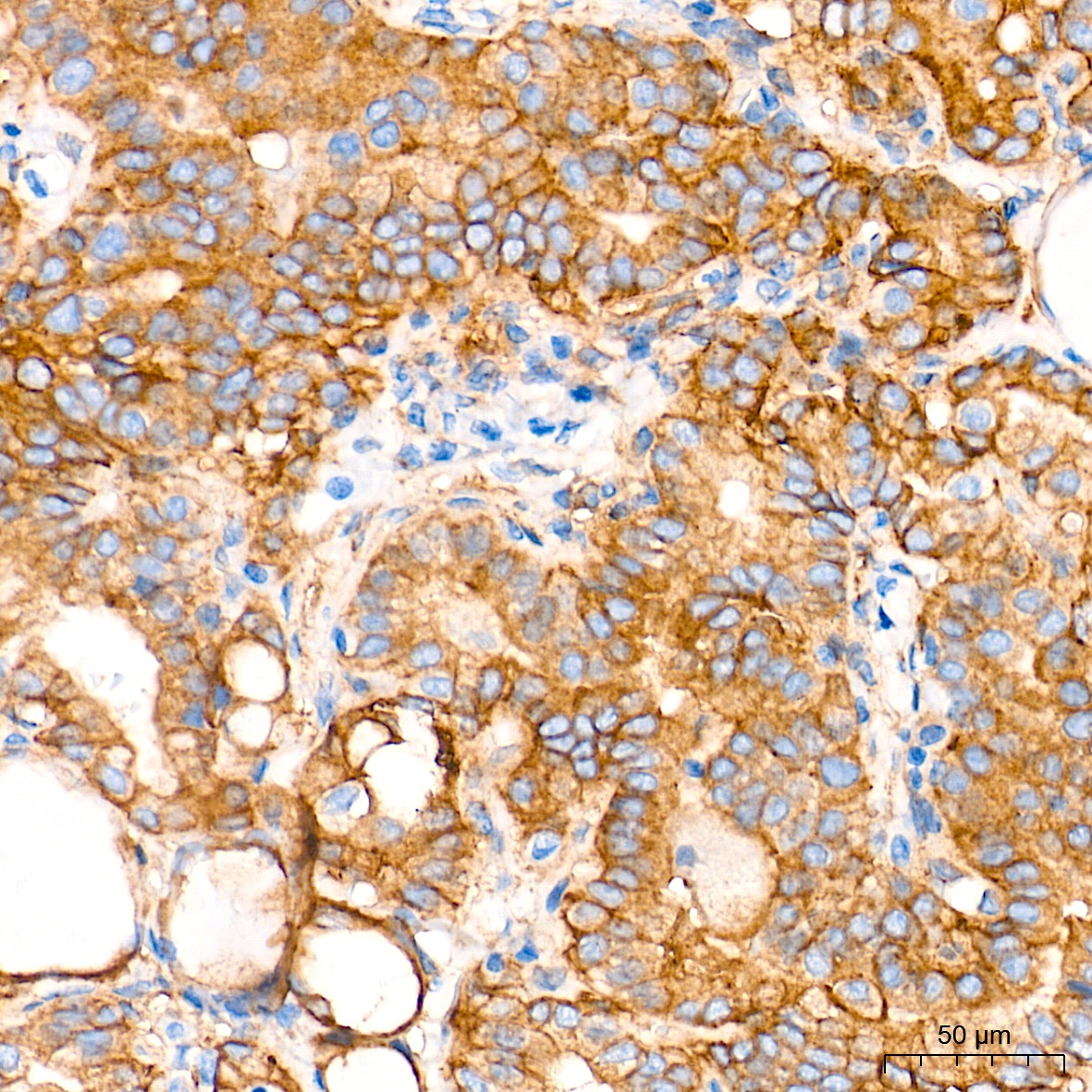
Immunohistochemistry analysis of paraffin-embedded Human spleen tissue using Integrin alpha V (ITGAV/CD51) Rabbit mAb (CAB19071) at a dilution of 1:2000 (40x lens). High pressure antigen retrieval performed with 0.01M Tris-EDTA Buffer (pH 9.0) prior to IHC staining.
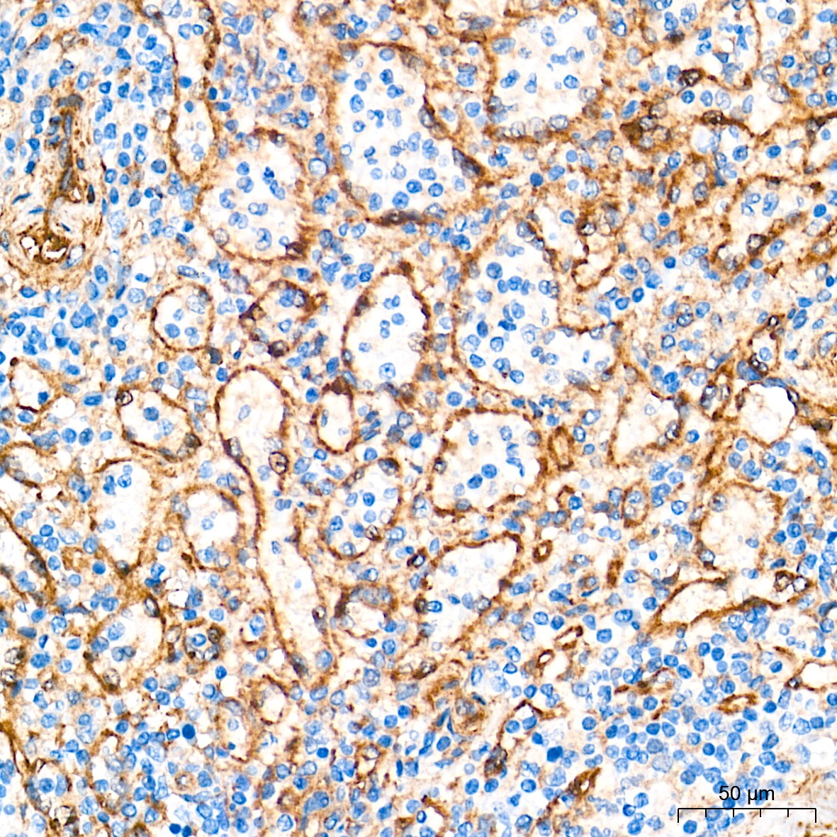
Immunohistochemistry analysis of paraffin-embedded Human thyroid cancer tissue using Integrin alpha V (ITGAV/CD51) Rabbit mAb (CAB19071) at a dilution of 1:2000 (40x lens). High pressure antigen retrieval performed with 0.01M Tris-EDTA Buffer (pH 9.0) prior to IHC staining.
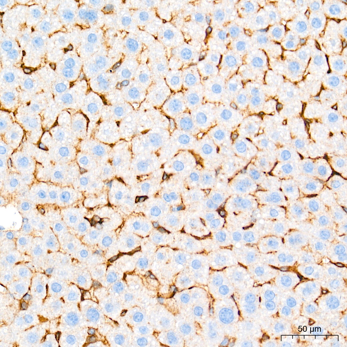
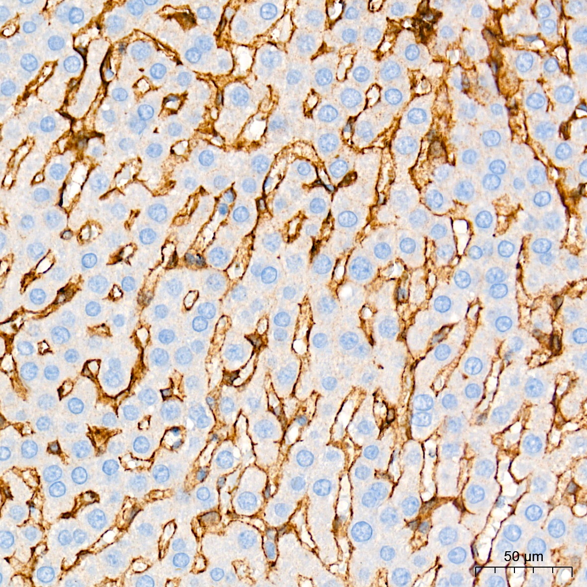
Immunohistochemistry analysis of paraffin-embedded Mouse liver tissue using Integrin alpha V (ITGAV/CD51) Rabbit mAb (CAB19071) at a dilution of 1:2000 (40x lens). High pressure antigen retrieval performed with 0.01M Tris-EDTA Buffer (pH 9.0) prior to IHC staining.
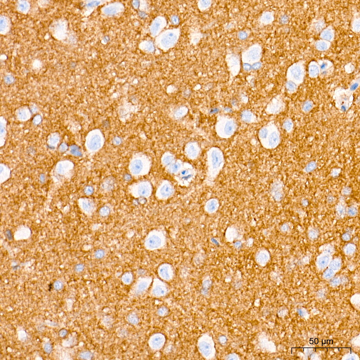
Immunohistochemistry analysis of paraffin-embedded Rat brain tissue using Integrin alpha V (ITGAV/CD51) Rabbit mAb (CAB19071) at a dilution of 1:2000 (40x lens). High pressure antigen retrieval performed with 0.01M Tris-EDTA Buffer (pH 9.0) prior to IHC staining.
Immunohistochemistry analysis of paraffin-embedded Rat liver tissue using Integrin alpha V (ITGAV/CD51) Rabbit mAb (CAB19071) at a dilution of 1:2000 (40x lens). High pressure antigen retrieval performed with 0.01M Tris-EDTA Buffer (pH 9.0) prior to IHC staining.
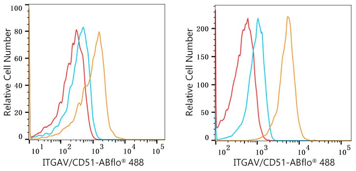
Flow cytometry:1X10^6 Daudi cells (negative control,left) and HUVEC cells (right) were intracellularly-stained with Integrin alpha V (ITGAV/CD51) Rabbit mAb(CAB19071, 2.5 μg/mL,orange line) or Rabbit IgG isotype control (AC042, 2.5 μg/mL,blue line),followed by FITC conjugated goat anti-rabbit pAb(1:200 dilution) staining. Non-fluorescently stained cells were used as blank control (red line).
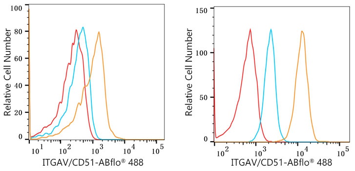
Flow cytometry:1X10^6 Daudi cells cells (negative control,left) and BEWO cells (right) were intracellularly-stained with Integrin alpha V (ITGAV/CD51) Rabbit mAb(CAB19071, 2.5 μg/mL,orange line) or Rabbit IgG isotype control (AC042, 2.5 μg/mL,blue line),followed by FITC conjugated goat anti-rabbit pAb(1:200 dilution) staining. Non-fluorescently stained cells were used as blank control (red line).
Flow cytometry:1X10^6 Daudi cells (negative control,left) and U-251MG cells (right) were intracellularly-stained with Integrin alpha V (ITGAV/CD51) Rabbit mAb(CAB19071, 2.5 μg/mL,orange line) or Rabbit IgG isotype control (AC042, 2.5 μg/mL,blue line),followed by FITC conjugated goat anti-rabbit pAb(1:200 dilution) staining. Non-fluorescently stained cells were used as blank control (red line).


 ELISA Kit (CHEB0059)")


