The CLU Antibody (CAB13479) is a high-quality antibody developed for reliable detection and analysis of target proteins. This antibody, raised in rabbits, is highly sensitive and specific to human samples, making it ideal for use in various applications such as Western blotting and immunohistochemistry.Clusterin, also known as apolipoprotein J, is a versatile protein that has been implicated in several disease processes, including cancer, neurodegenerative disorders, and cardiovascular diseases.
This antibody is validated for use in WB, IHC-P, IF/ICC, ELISA applications and has demonstrated reactivity against Human, Mouse, Rat samples.
Product Name:
CLU Antibody
SKU:
CAB13479
Size:
20μL, 100μL
Reactivity:
Human, Mouse, Rat
Conjugate:
Unconjugated
Immunogen:
Recombinant protein (or fragment).This information is considered to be commercially sensitive.
The protein encoded by this gene is a secreted chaperone that can under some stress conditions also be found in the cell cytosol. It has been suggested to be involved in several basic biological events such as cell death, tumor progression, and neurodegenerative disorders. Alternate splicing results in both coding and non-coding variants.
Purification Method
Affinity purification
Gene ID
1191
RRID
AB_2760341
Buffer Information
Store at -20℃. Avoid freeze / thaw cycles. Buffer: PBS containing 50% glycerol, preserved with proclin300 or sodium azide, pH 7.3.
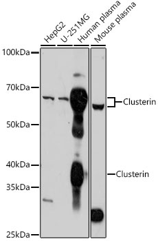
Western blot analysis of various lysates using Clusterin Rabbit pAb (CAB13479) at 1:1000 dilution. Secondary antibody: HRP-conjugated Goat anti-Rabbit IgG (H+L) (CABS014) at 1:10000 dilution. Lysates/proteins: 25μg per lane. Blocking buffer: 3% nonfat dry milk in TBST. Detection: ECL Basic Kit (AbGn00020). Exposure time: 30s.
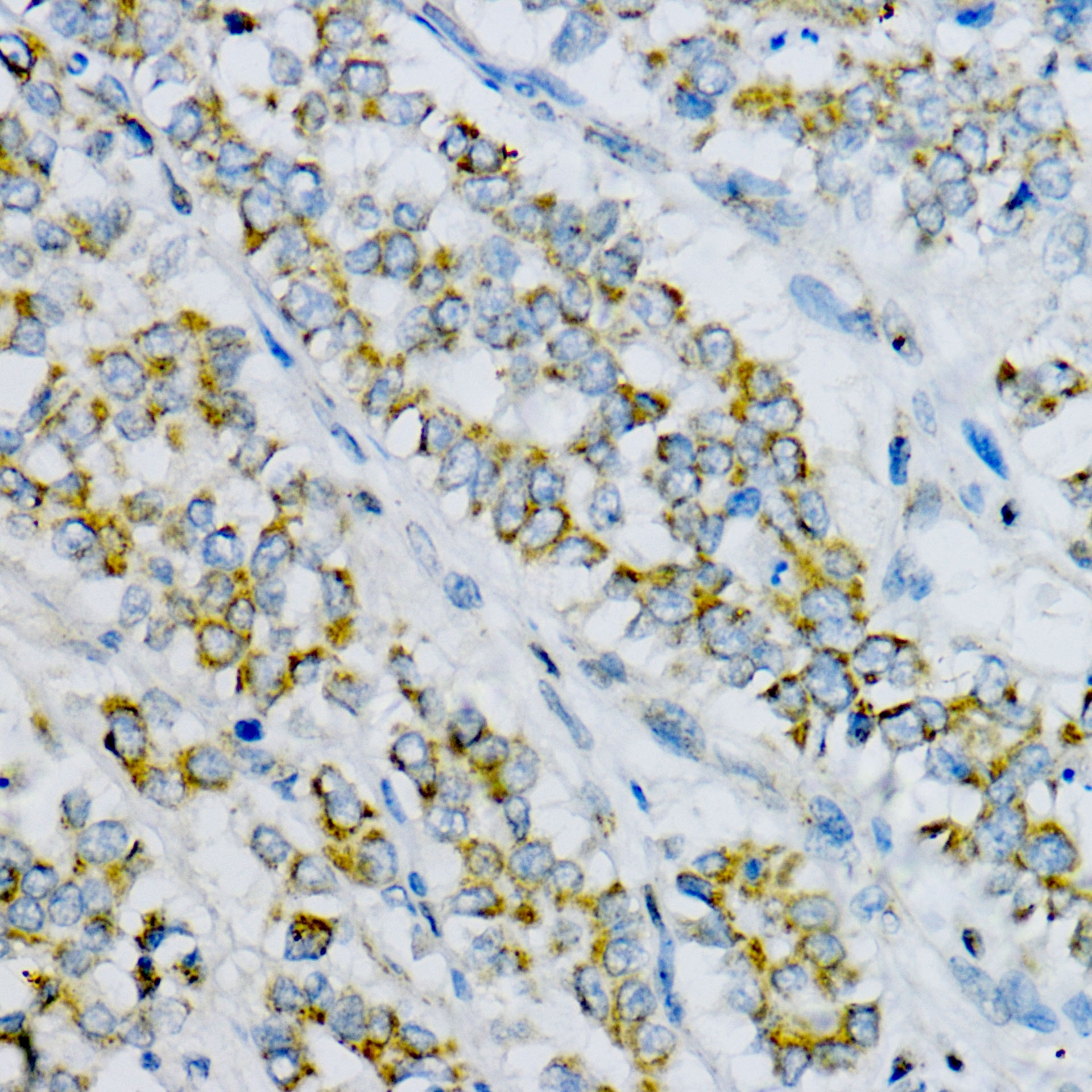
Immunohistochemistry analysis of paraffin-embedded Human colon cancer tissue using Clusterin Rabbit pAb (CAB13479) at a dilution of 1:100 (40x lens). High pressure antigen retrieval performed with 0.01M Citrate Buffer(pH 6.0) prior to IHC staining.
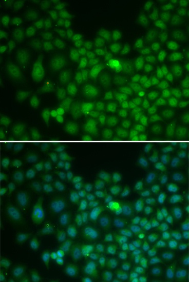
Immunofluorescence analysis of U2OS cells using Clusterin Rabbit pAb (CAB13479). Secondary antibody: Cy3-conjugated Goat anti-Rabbit IgG (H+L) (CABS007) at 1:500 dilution. Blue: DAPI for nuclear staining.


")

