The CNBP Antibody (CAB15110) is a high-quality antibody developed for reliable detection and analysis of target proteins. This antibody, produced in rabbits, is highly specific for detecting CNBP in human samples and has been validated for use in Western blot applications.CNBP is a multifunctional protein involved in diverse biological processes including transcriptional regulation, RNA processing, and telomere maintenance. Its versatile functions make it a key target for research in fields such as genetics, molecular biology, and developmental biology. The CNBP Polyclonal Antibody enables accurate detection and analysis of CNBP levels in different cell types, providing valuable insights for studies on gene expression regulation and nucleic acid metabolism.
This antibody is validated for use in WB, IHC-P, IF/ICC, ELISA applications and has demonstrated reactivity against Human, Mouse, Rat samples.
Product Name:
CNBP Antibody
SKU:
CAB15110
Size:
20μL, 100μL
Reactivity:
Human, Mouse, Rat
Conjugate:
Unconjugated
Immunogen:
Synthetic peptide. This information is considered to be commercially sensitive.
Sequence:
CGRG GHIA KDCK EPKR EREQ CCYN CGKP GHLA RDCD HADE QKCY SCGE FGHI QKDC TKVK CYRC GETG HVAI NCSK TSEV NCYR CGES GHLA RECT IEAT A
Tested Applications:
WBIHC-PIF/ICCELISA
Recommended Dilution:
WB
1:500 - 1:1000
IHC-P
1:50 - 1:200
IF/ICC
1:50 - 1:200
ELISA
Recommended starting concentration is 1 μg/mL. Please optimize the concentration based on your specific assay requirements.
Synonyms:
DM2, ZNF9, CNBP1, PROMM, RNF163, ZCCHC22, CNBP
Positive Sample:
HeLa, 293F, NIH/3T3
Cellular Localization:
Nucleus, Cytoplasm, Endoplasmic Reticulum.
Calculated MW:
19kDa
Observed MW:
20kDa
This gene encodes a nucleic-acid binding protein with seven zinc-finger domains. The protein has a preference for binding single stranded DNA and RNA. The protein functions in cap-independent translation of ornithine decarboxylase mRNA, and may also function in sterol-mediated transcriptional regulation. A CCTG expansion from <30 repeats to 75-11000 repeats in the first intron of this gene results in myotonic dystrophy type 2. Multiple transcript variants encoding different isoforms have been found for this gene.
Purification Method
Affinity purification
Gene ID
7555
RRID
AB_2761995
Buffer Information
Store at -20℃. Avoid freeze / thaw cycles. Buffer: PBS containing 50% glycerol, preserved with proclin300 or sodium azide, pH 7.3.
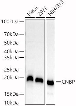
Western blot analysis of various lysates, using CNBP Rabbit pAb (CAB15110) at 1:800 dilution. Secondary antibody: HRP-conjugated Goat anti-Rabbit IgG (H+L) (CABS014) at 1:10000 dilution. Lysates/proteins: 25μg per lane. Blocking buffer: 3% nonfat dry milk in TBST. Detection: ECL Basic Kit (AbGn00020). Exposure time: 60s.
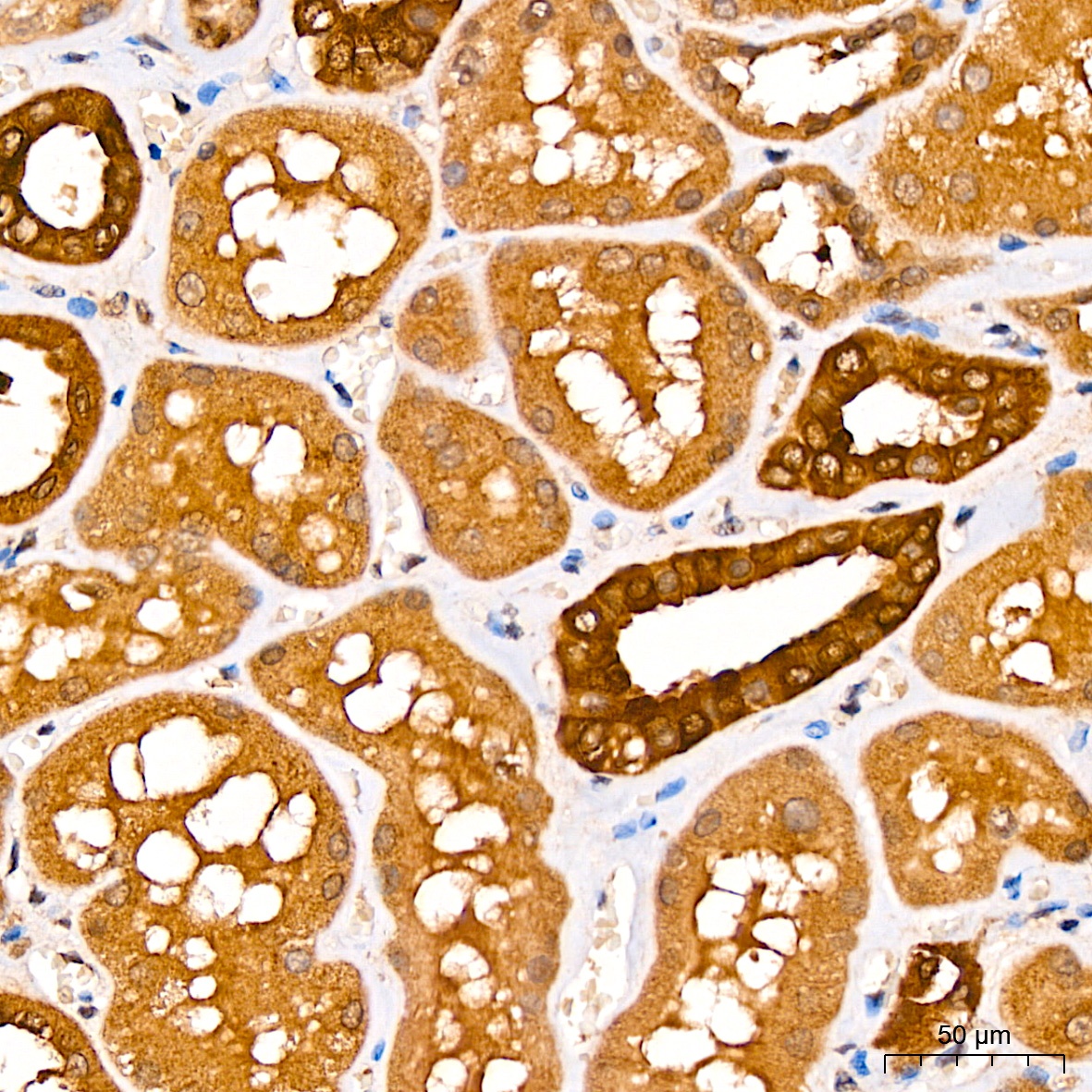
Immunohistochemistry analysis of paraffin-embedded Human kidney tissue using CNBP Rabbit pAb (CAB15110) at a dilution of 1:100 (40x lens). High pressure antigen retrieval was performed with 0.01 M citrate buffer (pH 6.0) prior to IHC staining.
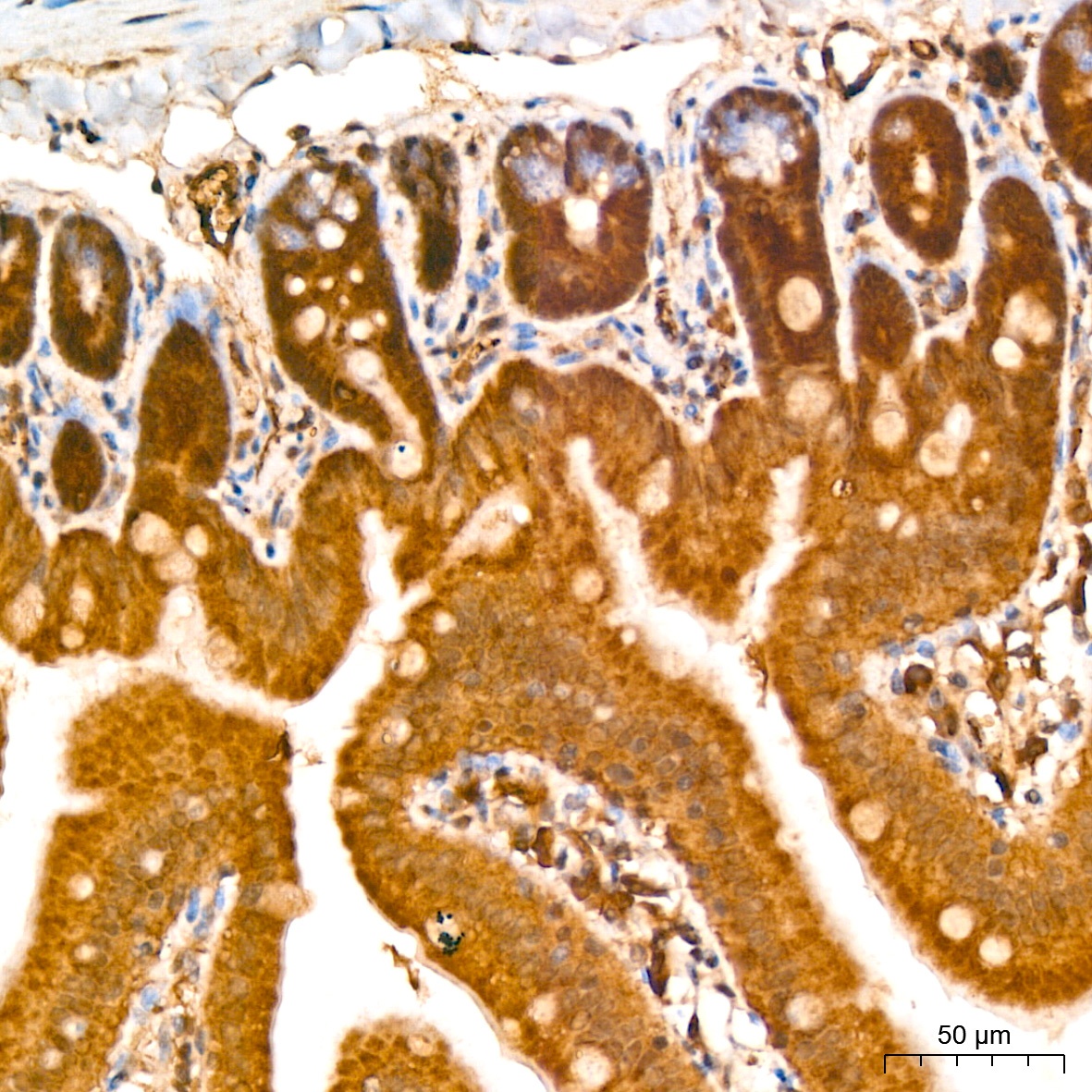
Immunohistochemistry analysis of paraffin-embedded Mouse colon tissue using CNBP Rabbit pAb (CAB15110) at a dilution of 1:100 (40x lens). High pressure antigen retrieval was performed with 0.01 M citrate buffer (pH 6.0) prior to IHC staining.
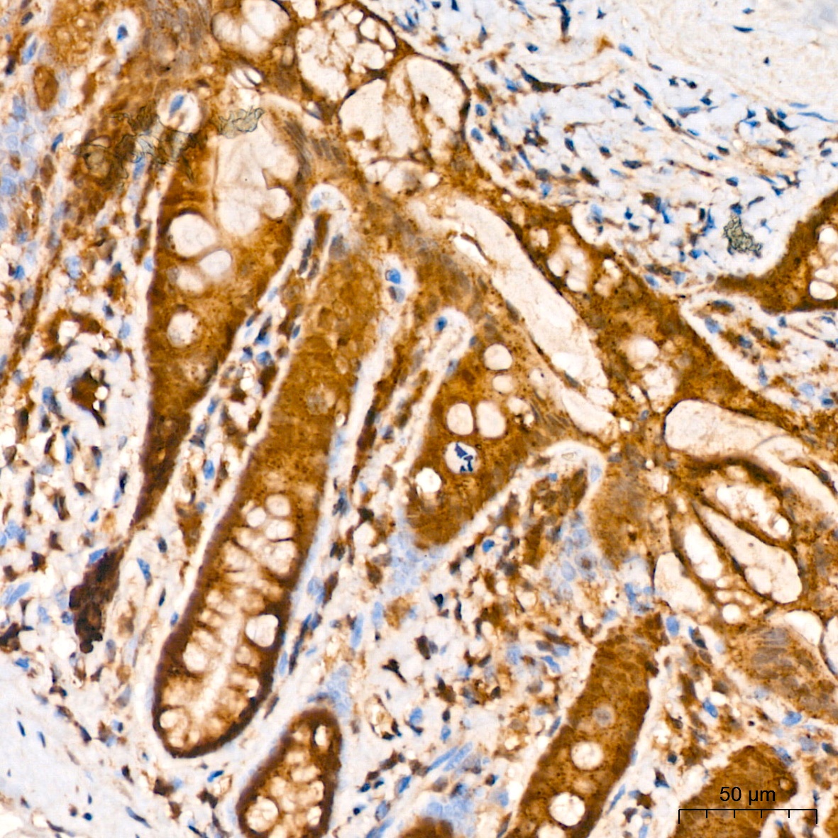
Immunohistochemistry analysis of paraffin-embedded Rat colon tissue using CNBP Rabbit pAb (CAB15110) at a dilution of 1:100 (40x lens). High pressure antigen retrieval was performed with 0.01 M citrate buffer (pH 6.0) prior to IHC staining.