The COPG2 Antibody (CAB15817) is a high-quality antibody developed for reliable detection and analysis of target proteins. This antibody, developed through immunization of rabbits, is highly specific and reactive with human samples, making it an excellent choice for Western blot analysis. By binding to the COPG2 protein, researchers can accurately detect and analyze its expression in various cell types, providing insights into its role in cellular processes.COPG2 is known to be essential for proper Golgi structure and function, and dysregulation of COPG2 has been implicated in various diseases, including cancer and neurodegenerative disorders.
This antibody is validated for use in WB, IHC-P, IF/ICC, ELISA applications and has demonstrated reactivity against Human, Mouse, Rat samples.
Product Name:
COPG2 Antibody
SKU:
CAB15817
Size:
20μL, 100μL
Reactivity:
Human, Mouse, Rat
Conjugate:
Unconjugated
Immunogen:
Recombinant protein (or fragment).This information is considered to be commercially sensitive.
Predicted to enable structural molecule activity. Involved in intra-Golgi vesicle-mediated transport. Part of COPI vesicle coat.
Purification Method
Affinity purification
Gene ID
26958
RRID
AB_2763240
Buffer Information
Store at -20℃. Avoid freeze / thaw cycles. Buffer: PBS with 0.01% thimerosal,50% glycerol,pH7.3.
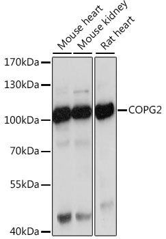
Western blot analysis of various lysates using COPG2 Rabbit pAb (CAB15817) at 1:1000 dilution. Secondary antibody: HRP-conjugated Goat anti-Rabbit IgG (H+L) (CABS014) at 1:10000 dilution. Lysates/proteins: 25μg per lane. Blocking buffer: 3% nonfat dry milk in TBST. Detection: ECL Basic Kit (AbGn00020). Exposure time: 30s.
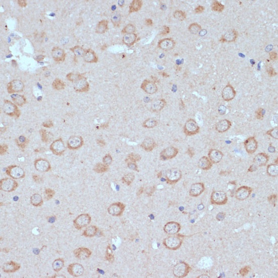
Immunohistochemistry analysis of paraffin-embedded Mouse brain using COPG2 Rabbit pAb (CAB15817) at dilution of 1:100 (40x lens). Microwave antigen retrieval performed with 0.01M PBS Buffer (pH 7.2) prior to IHC staining.