The Cortactin Monoclonal Antibody (CAB9518) is a high-quality antibody developed for reliable detection and analysis of target proteins. This gene is overexpressed in breast cancer and squamous cell carcinomas of the head and neck. The encoded protein is localized in the cytoplasm and in areas of the cell-substratum contacts. This gene has two roles: (1) regulating the interactions between components of adherens-type junctions and (2) organizing the cytoskeleton and cell adhesion structures of epithelia and carcinoma cells. During apoptosis, the encoded protein is degraded in a caspase-dependent manner. The aberrant regulation of this gene contributes to tumor cell invasion and metastasis. Three splice variants that encode different isoforms have been identified for this gene.
This antibody is validated for use in WB, IHC-P, IP, ELISA, IF-P applications and has demonstrated reactivity against Human, Mouse, Rat samples.
Product Name:
Cortactin Monoclonal Antibody
SKU:
CAB9518
Size:
100μL, 20μL
Reactivity:
Human, Mouse, Rat
Clone Number:
ARC1613
Conjugate:
Unconjugated
Immunogen:
Synthetic peptide. This information is considered to be commercially sensitive.
Tested Applications:
WBIHC-PIPELISAIF-P
Recommended Dilution:
WB
1:1000 - 1:6000
IP
0.5μg-4μg antibody for 400μg-600μg extracts of whole cells
IF-P
1:200 - 1:800
IHC-P
1:200 - 1:800
ELISA
Recommended starting concentration is 1 μg/mL. Please optimize the concentration based on your specific assay requirements.
This gene is overexpressed in breast cancer and squamous cell carcinomas of the head and neck. The encoded protein is localized in the cytoplasm and in areas of the cell-substratum contacts. This gene has two roles: (1) regulating the interactions between components of adherens-type junctions and (2) organizing the cytoskeleton and cell adhesion structures of epithelia and carcinoma cells. During apoptosis, the encoded protein is degraded in a caspase-dependent manner. The aberrant regulation of this gene contributes to tumor cell invasion and metastasis. Three splice variants that encode different isoforms have been identified for this gene.
Purification Method
Affinity purification
Gene ID
2017
RRID
AB_2863711
Buffer Information
Store at -20℃. Avoid freeze / thaw cycles. Buffer: PBS containing 50% glycerol and 0.05% BSA, preserved with proclin300 or sodium azide, pH 7.3.
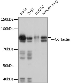
Western blot analysis of various lysates using Cortactin Rabbit mAb (CAB9518) at 1:1000 dilution. Secondary antibody: HRP-conjugated Goat anti-Rabbit IgG (H+L) (AS014) at 1:10000 dilution. Lysates/proteins: 25μg per lane. Blocking buffer: 3% nonfat dry milk in TBST. Detection: ECL Basic Kit (AbGn00020). Exposure time: 3s.
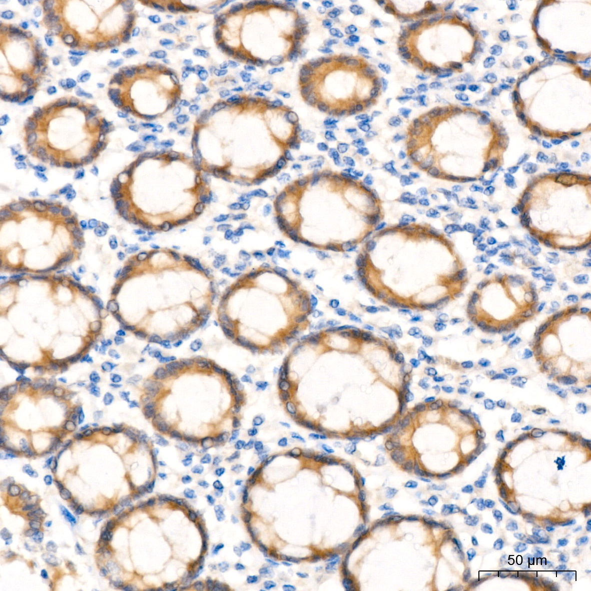
Immunohistochemistry analysis of paraffin-embedded Human colon tissue using Cortactin Rabbit mAb (CAB9518) at a dilution of 1:200 (40x lens). High pressure antigen retrieval performed with 0.01M Tris-EDTA Buffer (pH 9.0) prior to IHC staining.
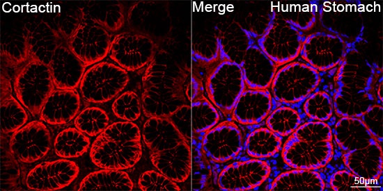
Confocal imaging of paraffin-embedded Human stomach tissue using Cortactin Rabbit mAb (CAB9518, dilution 1:200) followed by a further incubation with Cy3 Goat Anti-Rabbit IgG (H+L) (AS007, dilution 1:500) (Red). DAPI was used for nuclear staining (Blue). Objective: 40x. Perform high pressure antigen retrieval with 0.01 M citrate buffer (pH 6.0) prior to IF staining.
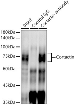
Immunoprecipitation analysis of 600 μg extracts of Mouse lung cells using 3 μg Cortactin antibody (CAB9518). Western blot was performed from the immunoprecipitate using Cortactin antibody (CAB9518) at a dilution of 1:1000.