The COX5A Polyclonal Antibody (CAB24500) is a high-quality antibody developed for reliable detection and analysis of target proteins. This antibody, generated in rabbits, is highly specific to human samples and has been validated for use in Western blot applications. By binding to the COX5A protein, researchers can accurately detect and analyze its expression in various cell types, making it a valuable tool for studies in mitochondrial biology and respiratory chain function.COX5A is known to play a crucial role in mitochondrial energy production and cellular respiration. Mutations or dysregulation of this protein have been linked to various mitochondrial diseases and disorders.
This antibody is validated for use in WB, IHC-P, ELISA applications and has demonstrated reactivity against Human, Mouse, Rat samples.
Product Name:
COX5A Polyclonal Antibody
SKU:
CAB24500
Size:
20μL, 100μL
Reactivity:
Human, Mouse, Rat
Conjugate:
Unconjugated
Immunogen:
Recombinant protein (or fragment).This information is considered to be commercially sensitive.
Recommended starting concentration is 1 μg/mL. Please optimize the concentration based on your specific assay requirements.
Synonyms:
COX5A, COX, COX-VA, VA, cytochrome c oxidase subunit 5A, 5A
Positive Sample:
HeLa, Mouse brain, Mouse heart, Rat brain
Cellular Localization:
Mitochondrion Inner Membrane.
Calculated MW:
16kDa
Observed MW:
15kDa
Cytochrome c oxidase (COX) is the terminal enzyme of the mitochondrial respiratory chain. It is a multi-subunit enzyme complex that couples the transfer of electrons from cytochrome c to molecular oxygen and contributes to a proton electrochemical gradient across the inner mitochondrial membrane. The complex consists of 13 mitochondrial- and nuclear-encoded subunits. The mitochondrially-encoded subunits perform the electron transfer of proton pumping activities. The functions of the nuclear-encoded subunits are unknown but they may play a role in the regulation and assembly of the complex. This gene encodes the nuclear-encoded subunit Va of the human mitochondrial respiratory chain enzyme. A pseudogene COX5AP1 has been found in chromosome 14q22.
Purification Method
Affinity purification
Gene ID
9377
Buffer Information
Store at -20℃. Avoid freeze / thaw cycles. Buffer: PBS containing 50% glycerol, preserved with proclin300 or sodium azide, pH 7.3.
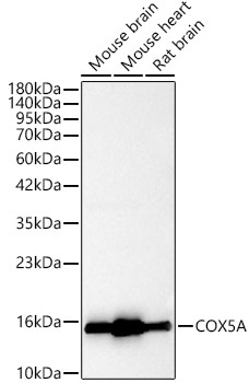
Western blot analysis of various lysates, using [KO Validated] COX5A Rabbit pAb (CAB24500) at 1:2000 dilution. Secondary antibody: HRP-conjugated Goat anti-Rabbit IgG (H+L) (CABS014) at 1:10000 dilution. Lysates/proteins: 25μg per lane. Blocking buffer: 3% nonfat dry milk in TBST. Detection: ECL Basic Kit (AbGn00020). Exposure time: 60s.
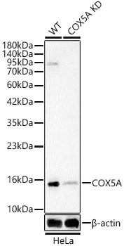
Western blot analysis of lysates from wild type (WT) and COX5A knockdown (KD) HeLa(KD) cells, using [KO Validated] COX5A Rabbit pAb (CAB24500) at 1:2000 dilution. Secondary antibody: HRP-conjugated Goat anti-Rabbit IgG (H+L) (CABS014) at 1:10000 dilution. Lysates/proteins: 25ug per lane. Blocking buffer: 3% nonfat dry milk in TBST. Detection: ECL Basic Kit (AbGn00020). Exposure time: 60s.
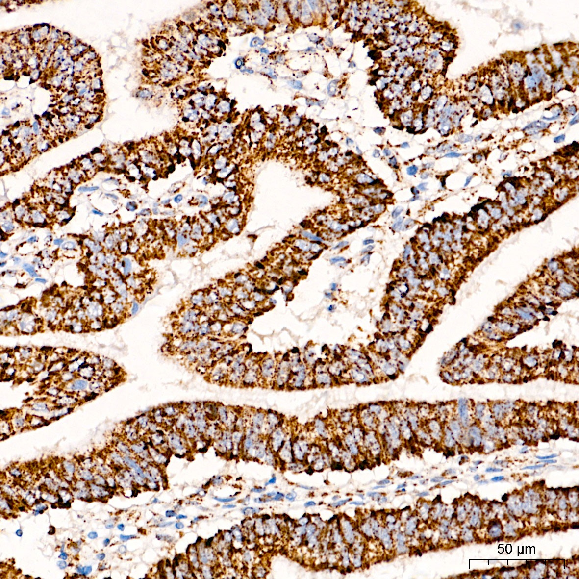
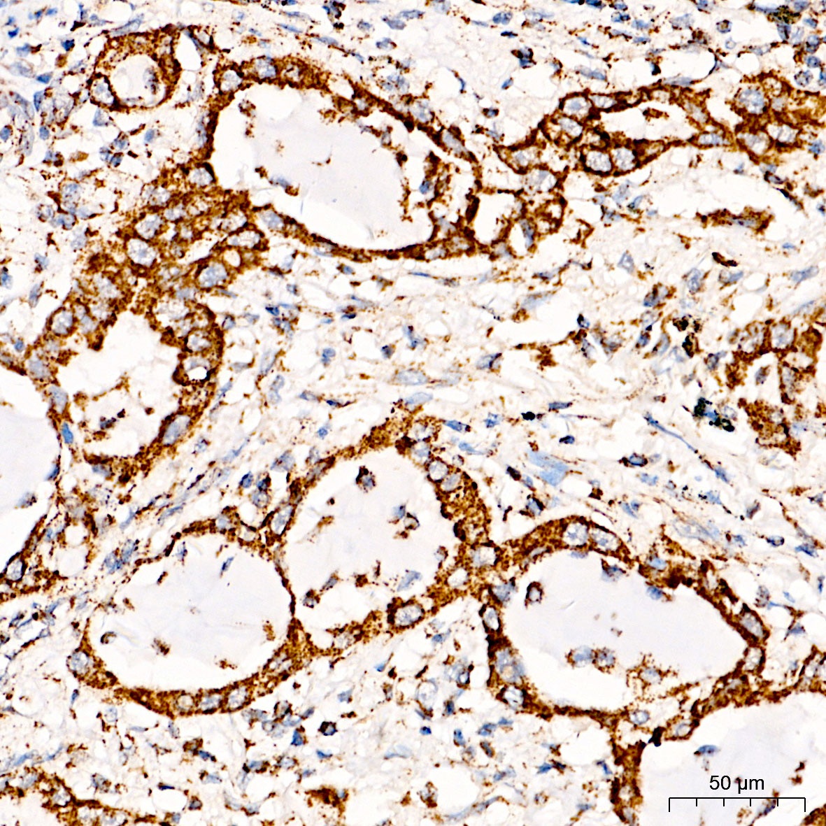
Immunohistochemistry analysis of paraffin-embedded Human colon carcinoma tissue using [KO Validated] COX5A Rabbit pAb (CAB24500) at a dilution of 1:100 (40x lens). High pressure antigen retrieval was performed with 0.01 M citrate buffer (pH 6.0) prior to IHC staining.
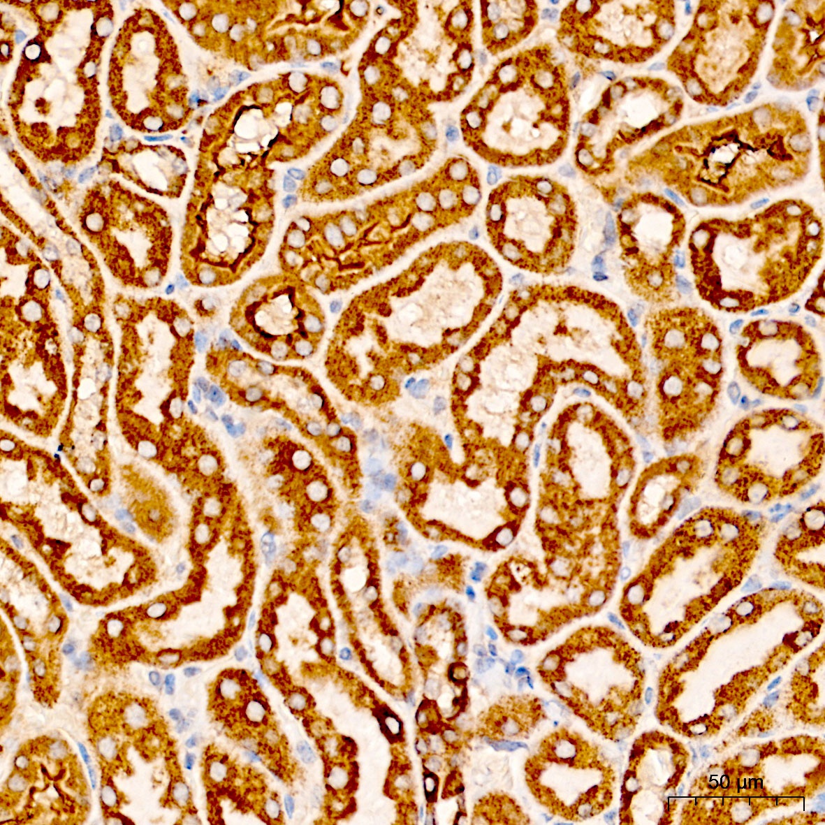
Immunohistochemistry analysis of paraffin-embedded Mouse kidney tissue using [KO Validated] COX5A Rabbit pAb (CAB24500) at a dilution of 1:100 (40x lens). High pressure antigen retrieval was performed with 0.01 M citrate buffer (pH 6.0) prior to IHC staining.
Immunohistochemistry analysis of paraffin-embedded Human lung cancer tissue using [KO Validated] COX5A Rabbit pAb (CAB24500) at a dilution of 1:100 (40x lens). High pressure antigen retrieval was performed with 0.01 M citrate buffer (pH 6.0) prior to IHC staining.
![Western blot analysis of various lysates, using [KO Validated] COX5A Rabbit pAb (CAB24500) at 1:2000 dilution. Secondary antibody: HRP Goat Anti-Rabbit IgG (H+L) at 1:10000 dilution. Lysates/proteins: 25ug per lane. Blocking buffer: 3% nonfat dry milk in TBST.](https://cdn11.bigcommerce.com/s-h68l9z2lnx/images/stencil/1280x1280/products/224342/583604/A24500_N29296-4_WB_01__06975.1701346913.jpg?c=2?imbypass=on "Western blot analysis of various lysates, using [KO Validated] COX5A Rabbit pAb (CAB24500) at 1:2000 dilution. Secondary antibody: HRP Goat Anti-Rabbit IgG (H+L) at 1:10000 dilution. Lysates/proteins: 25ug per lane. Blocking buffer: 3% nonfat dry milk in TBST.")
![Western blot analysis of various lysates, using [KO Validated] COX5A Rabbit pAb (CAB24500) at 1:2000 dilution. Secondary antibody: HRP Goat Anti-Rabbit IgG (H+L) at 1:10000 dilution. Lysates/proteins: 25ug per lane. Blocking buffer: 3% nonfat dry milk in TBST.](https://cdn11.bigcommerce.com/s-h68l9z2lnx/images/stencil/608x608/products/224342/583604/A24500_N29296-4_WB_01__06975.1701346913.jpg?c=2 "Western blot analysis of various lysates, using [KO Validated] COX5A Rabbit pAb (CAB24500) at 1:2000 dilution. Secondary antibody: HRP Goat Anti-Rabbit IgG (H+L) at 1:10000 dilution. Lysates/proteins: 25ug per lane. Blocking buffer: 3% nonfat dry milk in TBST.")


")
 at 1:10000 dilution. Lysates/proteins: 25ug per lane. Blocking buffer: 3% nonfat dry milk in TBST. Detection: ECL Basic Kit. Exposure time: 1s.")
