The COX6A1 Antibody (CAB3798) is a high-quality antibody developed for reliable detection and analysis of target proteins. This antibody, raised in rabbits, is highly specific to human samples and has been validated for use in Western blot applications. By binding to the COX6A1 protein, this antibody enables accurate detection and analysis in a variety of cell types, making it ideal for studies in biochemistry and mitochondrial research.COX6A1 is a subunit of the cytochrome c oxidase complex, which plays a crucial role in the electron transport chain and ATP production in mitochondria.
This antibody is validated for use in WB, IHC-P, IF/ICC, ELISA applications and has demonstrated reactivity against Human, Mouse, Rat samples.
Product Name:
COX6A1 Antibody
SKU:
CAB3798
Size:
20μL, 100μL
Reactivity:
Human, Mouse, Rat
Conjugate:
Unconjugated
Immunogen:
Recombinant protein (or fragment).This information is considered to be commercially sensitive.
Recommended starting concentration is 1 μg/mL. Please optimize the concentration based on your specific assay requirements.
Synonyms:
COX6A, CMTRID, COX6AL, COX6A1
Positive Sample:
A-549, MCF7, Mouse kidney, Mouse heart, Mouse liver, Rat liver, Rat brain
Cellular Localization:
Mitochondrion Inner Membrane.
Calculated MW:
12kDa
Observed MW:
12kDa
Cytochrome c oxidase (COX), the terminal enzyme of the mitochondrial respiratory chain, catalyzes the electron transfer from reduced cytochrome c to oxygen. It is a heteromeric complex consisting of 3 catalytic subunits encoded by mitochondrial genes and multiple structural subunits encoded by nuclear genes. The mitochondrially-encoded subunits function in the electron transfer and the nuclear-encoded subunits may function in the regulation and assembly of the complex. This nuclear gene encodes polypeptide 1 (liver isoform) of subunit VIa, and polypeptide 1 is found in all non-muscle tissues. Polypeptide 2 (heart/muscle isoform) of subunit VIa is encoded by a different gene, and is present only in striated muscles. These two polypeptides share 66% amino acid sequence identity. It has been reported that there may be several pseudogenes on chromosomes 1, 6, 7q21, 7q31-32 and 12. However, only one pseudogene (COX6A1P) on chromosome 1p31.1 has been documented.
Purification Method
Affinity purification
Gene ID
1337
RRID
AB_2765311
Buffer Information
Store at -20℃. Avoid freeze / thaw cycles. Buffer: PBS containing 50% glycerol, preserved with proclin300 or sodium azide, pH 7.3.
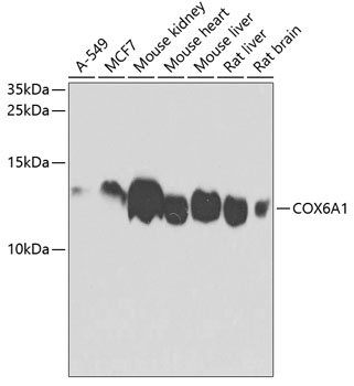
Western blot analysis of various lysates using COX6A1 Rabbit pAb (CAB3798) at 1:1000 dilution. Secondary antibody: HRP-conjugated Goat anti-Rabbit IgG (H+L) (CABS014) at 1:10000 dilution. Lysates/proteins: 25μg per lane. Blocking buffer: 3% nonfat dry milk in TBST. Detection: ECL Basic Kit (AbGn00020). Exposure time: 60s.
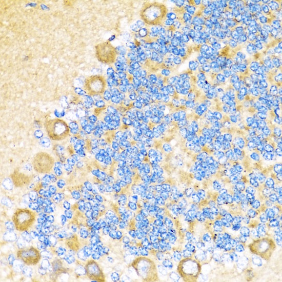
Immunohistochemistry analysis of paraffin-embedded Rat cerebellum using COX6A1 Rabbit pAb (CAB3798) at dilution of 1:100 (40x lens). Microwave antigen retrieval performed with 0.01M PBS Buffer (pH 7.2) prior to IHC staining.
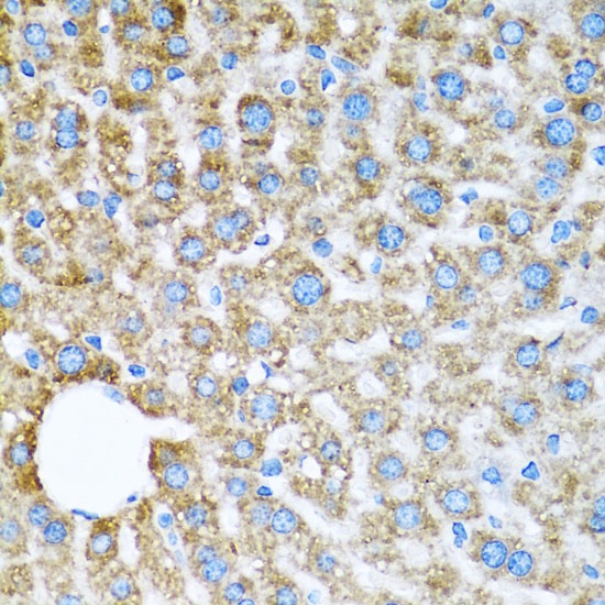
Immunohistochemistry analysis of paraffin-embedded Mouse liver using COX6A1 Rabbit pAb (CAB3798) at dilution of 1:100 (40x lens). Microwave antigen retrieval performed with 0.01M PBS Buffer (pH 7.2) prior to IHC staining.
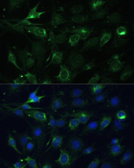
Immunofluorescence analysis of C6 cells using COX6A1 Rabbit pAb (CAB3798) at dilution of 1:100 (40x lens). Secondary antibody: Cy3-conjugated Goat anti-Rabbit IgG (H+L) (CABS007) at 1:500 dilution. Blue: DAPI for nuclear staining.
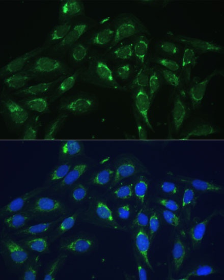
Immunofluorescence analysis of U-2 OS cells using COX6A1 Rabbit pAb (CAB3798) at dilution of 1:100 (40x lens). Secondary antibody: Cy3-conjugated Goat anti-Rabbit IgG (H+L) (CABS007) at 1:500 dilution. Blue: DAPI for nuclear staining.