The COX6C Antibody (CAB16250) is a high-quality antibody developed for reliable detection and analysis of target proteins. This antibody, derived from rabbits, is highly specific to human samples and has been validated for use in Western blot applications. By binding to the COX6C protein, this antibody allows for the detection and analysis of this important mitochondrial protein in various cell types.COX6C plays a critical role in the electron transport chain, facilitating the conversion of oxygen to water and ultimately producing ATP, the energy currency of the cell.
This antibody is validated for use in WB, IF/ICC, ELISA applications and has demonstrated reactivity against Mouse samples.
Product Name:
COX6C Antibody
SKU:
CAB16250
Size:
20μL, 100μL
Reactivity:
Mouse
Conjugate:
Unconjugated
Immunogen:
Synthetic peptide. This information is considered to be commercially sensitive.
Recommended starting concentration is 1 μg/mL. Please optimize the concentration based on your specific assay requirements.
Synonyms:
COX6C
Cellular Localization:
Mitochondrial Inner Membrane, Mitochondrial Respiratory Chain Complex Iv, Mitochondrion.
Calculated MW:
9kDa
Observed MW:
Refertofigures
Cytochrome c oxidase, the terminal enzyme of the mitochondrial respiratory chain, catalyzes the electron transfer from reduced cytochrome c to oxygen. It is a heteromeric complex consisting of 3 catalytic subunits encoded by mitochondrial genes and multiple structural subunits encoded by nuclear genes. The mitochondrially-encoded subunits function in electron transfer, and the nuclear-encoded subunits may be involved in the regulation and assembly of the complex. This nuclear gene encodes subunit VIc, which has 77% amino acid sequence identity with mouse subunit VIc. This gene is up-regulated in prostate cancer cells. A pseudogene has been found on chromosomes 16p12.
Purification Method
Affinity purification
Gene ID
1345
RRID
AB_2769017
Buffer Information
Store at -20℃. Avoid freeze / thaw cycles. Buffer: PBS with 0.01% thimerosal,50% glycerol,pH7.3.
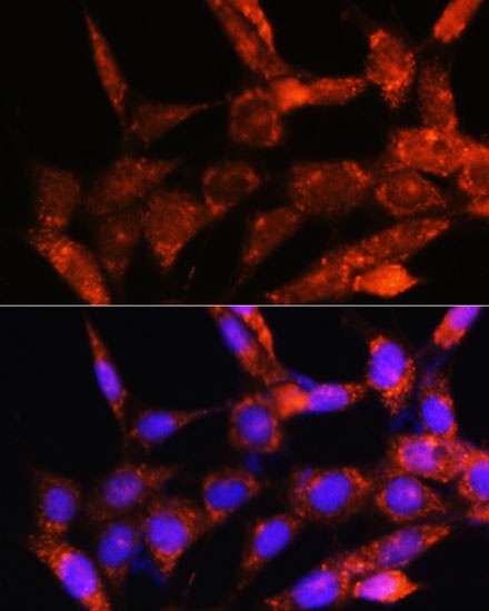
Immunofluorescence analysis of NIH/3T3 cells using COX6C Rabbit pAb (CAB16250) at dilution of 1:100. Secondary antibody: Cy3-conjugated Goat anti-Rabbit IgG (H+L) (CABS007) at 1:500 dilution. Blue: DAPI for nuclear staining.