The CPT1B Antibody (CAB6796) is a high-quality antibody developed for reliable detection and analysis of target proteins. This antibody, raised in rabbits, has high reactivity with human samples and is validated for use in Western blot applications. By targeting the CPT1B protein, researchers can detect and analyze this key enzyme in various cell types, making it ideal for studies in metabolism, obesity, and metabolic disorders.CPT1B is an essential enzyme in the regulation of fatty acid oxidation, making it a potential target for therapeutic interventions in diseases like obesity, diabetes, and cardiovascular disorders.
This antibody is validated for use in WB, IHC-P, ELISA, IF-P applications and has demonstrated reactivity against Human, Mouse, Rat samples.
Product Name:
CPT1B Antibody
SKU:
CAB6796
Size:
20μL, 100μL
Reactivity:
Human, Mouse, Rat
Conjugate:
Unconjugated
Immunogen:
Recombinant protein (or fragment).This information is considered to be commercially sensitive.
The protein encoded by this gene, a member of the carnitine/choline acetyltransferase family, is the rate-controlling enzyme of the long-chain fatty acid beta-oxidation pathway in muscle mitochondria. This enzyme is required for the net transport of long-chain fatty acyl-CoAs from the cytoplasm into the mitochondria. Multiple transcript variants encoding different isoforms have been found for this gene, and read-through transcripts are expressed from the upstream locus that include exons from this gene.
Purification Method
Affinity purification
Gene ID
1375
RRID
AB_2767379
Buffer Information
Store at -20℃. Avoid freeze / thaw cycles. Buffer: PBS containing 50% glycerol, preserved with proclin300 or sodium azide, pH 7.3.
Western blot analysis of lysates from Rat skeletal muscle, using CPT1B Rabbit pAb (CAB6796) at 1:500 dilution. Secondary antibody: HRP-conjugated Goat anti-Rabbit IgG (H+L) (CABS014) at 1:10000 dilution. Lysates/proteins: 25μg per lane. Blocking buffer: 3% nonfat dry milk in TBST. Detection: ECL Basic Kit (AbGn00020). Exposure time: 180s.
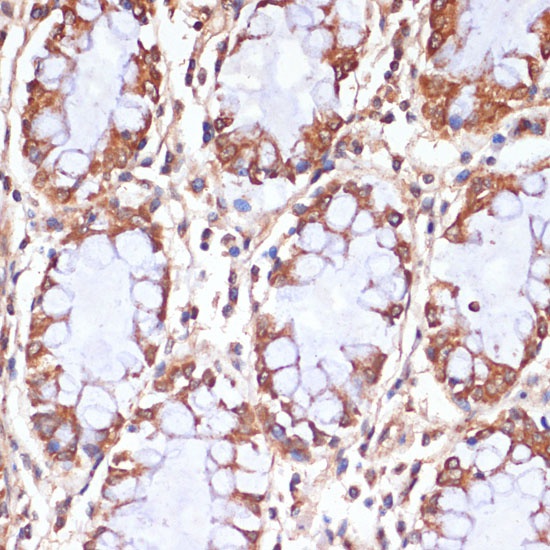
Immunohistochemistry analysis of paraffin-embedded Human colon using CPT1B Rabbit pAb (CAB6796) at dilution of 1:100 (40x lens). Microwave antigen retrieval performed with 0.01M PBS Buffer (pH 7.2) prior to IHC staining.
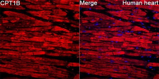
Immunofluorescence analysis of Human heart tissue using CPT1B Rabbit pAb (CAB6796) at a dilution of 1:100 (40x lens). Secondary antibody: Cy3-conjugated Goat anti-Rabbit IgG (H+L)(CABS007) at 1:500 dilution. Blue: DAPI for nuclear staining. High pressure antigen retrieval performed with 0.01M Citrate Buffer(pH 6.0) prior to IF staining.
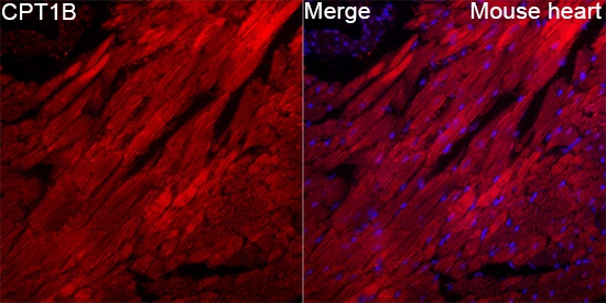
Immunofluorescence analysis of Mouse heart tissue using CPT1B Rabbit pAb (CAB6796) at a dilution of 1:100 (40x lens). Secondary antibody: Cy3-conjugated Goat anti-Rabbit IgG (H+L)(CABS007) at 1:500 dilution. Blue: DAPI for nuclear staining. High pressure antigen retrieval performed with 0.01M Citrate Buffer(pH 6.0) prior to IF staining.