The CRALBP Monoclonal Antibody (CAB9265) is a high-quality antibody developed for reliable detection and analysis of target proteins. CRALBP is a key protein involved in the visual cycle and retinoid metabolism, making it essential for vision and eye health.This antibody, produced in rabbits, is highly specific and reacts specifically with human samples, allowing for accurate and reliable results in research applications such as immunofluorescence and immunohistochemistry. By binding to CRALBP, this antibody enables the visualization and analysis of CRALBP expression in various tissues and cell types.The role of CRALBP in retinal function and metabolism makes it an important target for studies in vision research, ocular diseases, and retinal degenerative disorders.
This antibody is validated for use in WB, IHC-P, ELISA, IF-P applications and has demonstrated reactivity against Mouse, Rat samples.
Product Name:
CRALBP Monoclonal Antibody
SKU:
CAB9265
Size:
20μL, 100μL
Reactivity:
Mouse, Rat
Clone Number:
ARC1502
Conjugate:
Unconjugated
Immunogen:
Synthetic peptide. This information is considered to be commercially sensitive.
Sequence:
Email for sequence
Tested Applications:
WBIHC-PELISAIF-P
Recommended Dilution:
WB
1:1000 - 1:6000
IF-P
1:200 - 1:2000
IHC-P
1:100 - 1:1000
ELISA
Recommended starting concentration is 1 μg/mL. Please optimize the concentration based on your specific assay requirements.
Synonyms:
CRALBP
Positive Sample:
Mouse eye
Cellular Localization:
Cytoplasm.
Calculated MW:
36kDa
Observed MW:
36kDa
The protein encoded by this gene is a 36-kD water-soluble protein which carries 11-cis-retinaldehyde or 11-cis-retinal as physiologic ligands. It may be a functional component of the visual cycle. Mutations of this gene have been associated with severe rod-cone dystrophy, Bothnia dystrophy (nonsyndromic autosomal recessive retinitis pigmentosa) and retinitis punctata albescens.
Purification Method
Affinity purification
Gene ID
6017
RRID
AB_2863700
Buffer Information
Store at -20℃. Avoid freeze / thaw cycles. Buffer: PBS containing 50% glycerol and 0.05% BSA, preserved with proclin300 or sodium azide, pH 7.3.
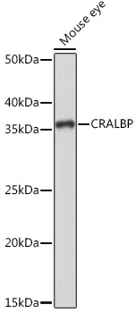
Western blot analysis of lysates from Mouse eye, using CRALBP Rabbit mAb (CAB9265) at 1:1000 dilution. Secondary antibody: HRP-conjugated Goat anti-Rabbit IgG (H+L) (CABS014) at 1:10000 dilution. Lysates/proteins: 25μg per lane. Blocking buffer: 3% nonfat dry milk in TBST. Detection: ECL Basic Kit (AbGn00020). Exposure time: 1s.
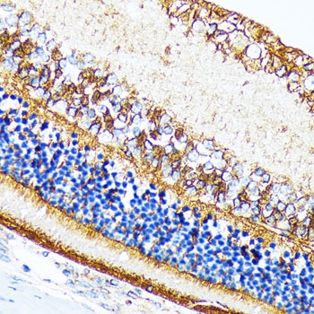
Immunohistochemistry analysis of paraffin-embedded Rat retina using CRALBP Rabbit mAb (CAB9265) at dilution of 1:100 (40x lens). Microwave antigen retrieval performed with 0.01M Tris/EDTA Buffer (pH 9.0) prior to IHC staining.
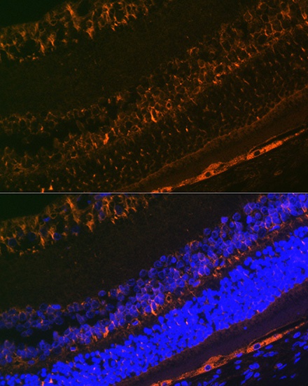
Immunofluorescence analysis of paraffin-embedded rat eye using CRALBP Rabbit mAb (CAB9265) at dilution of 1:100 (40x lens). Secondary antibody: Cy3-conjugated Goat anti-Rabbit IgG (H+L) (CABS007) at 1:500 dilution. Blue: DAPI for nuclear staining.
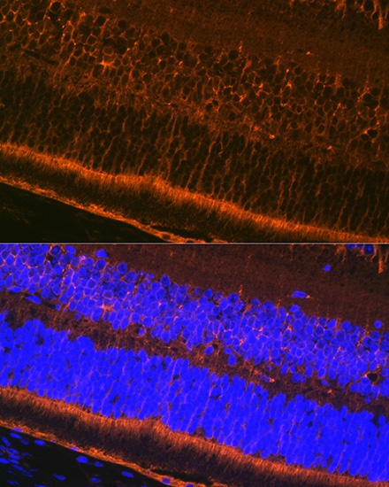
Immunofluorescence analysis of paraffin-embedded mouse eye using CRALBP Rabbit mAb (CAB9265) at dilution of 1:100 (40x lens). Secondary antibody: Cy3-conjugated Goat anti-Rabbit IgG (H+L) (CABS007) at 1:500 dilution. Blue: DAPI for nuclear staining.


 ELISA Kit (HUFI03087)")
 ELISA Kit (HUFI03337)")


