The CTCF Antibody (CAB13272) is a high-quality antibody developed for reliable detection and analysis of target proteins. This antibody, produced in rabbits, exhibits strong reactivity with human samples and has been validated for use in Western blot applications. By specifically binding to the CTCF protein, this antibody facilitates the detection and analysis of CTCF in various cell types, making it an essential component for studies in genetics, epigenetics, and cancer research.CTCF is a critical protein that plays a pivotal role in the regulation of gene expression by facilitating interactions between DNA sequences and influencing chromatin structure. Its involvement in various cellular processes, including DNA methylation, chromatin looping, and genomic stability, underscores its significance as a target for investigation in diverse fields of study.
This antibody is validated for use in WB, IHC-P, IF/ICC, IP, ChIP, ELISA applications and has demonstrated reactivity against Human, Mouse, Rat samples.
Product Name:
CTCF Antibody
SKU:
CAB13272
Size:
20μL, 100μL
Reactivity:
Human, Mouse, Rat
Conjugate:
Unconjugated
Immunogen:
Recombinant protein (or fragment).This information is considered to be commercially sensitive.
0.5μg-4μg antibody for 200μg-400μg extracts of whole cells
ELISA
Recommended starting concentration is 1 μg/mL. Please optimize the concentration based on your specific assay requirements.
ChIP
3μg antibody for 10μg-15μg of Chromatin
Synonyms:
MRD21, FAP108, CFAP108, CTCF
Positive Sample:
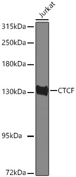
293T, Jurkat
Cellular Localization:
Chromosome, Nucleus, Centromere, Nucleoplasm.
Calculated MW:
83kDa
Observed MW:
140kDa
This gene is a member of the BORIS + CTCF gene family and encodes a transcriptional regulator protein with 11 highly conserved zinc finger (ZF) domains. This nuclear protein is able to use different combinations of the ZF domains to bind different DNA target sequences and proteins. Depending upon the context of the site, the protein can bind a histone acetyltransferase (HAT)-containing complex and function as a transcriptional activator or bind a histone deacetylase (HDAC)-containing complex and function as a transcriptional repressor. If the protein is bound to a transcriptional insulator element, it can block communication between enhancers and upstream promoters, thereby regulating imprinted expression. Mutations in this gene have been associated with invasive breast cancers, prostate cancers, and Wilms' tumors. Alternatively spliced transcript variants encoding different isoforms have been found for this gene.
Purification Method
Affinity purification
Gene ID
10664
RRID
AB_2760124
Buffer Information
Store at -20℃. Avoid freeze / thaw cycles. Buffer: PBS with 0.09% Sodium azide,50% glycerol,pH7.3.
Western blot analysis of lysates from Jurkat cells using CTCF Rabbit pAb (CAB13272) at 1:1000 dilution incubated overnight at 4℃. Secondary antibody: HRP-conjugated Goat anti-Rabbit IgG (H+L) (CABS014) at 1:10000 dilution. Lysates/proteins: 25 μg per lane. Blocking buffer: 3% nonfat dry milk in TBST. Detection: ECL Basic Kit (AbGn00020). Exposure time: 30 s.
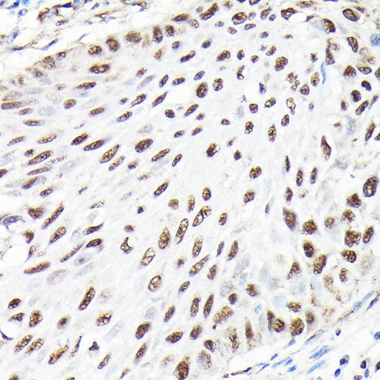
Immunohistochemistry analysis of paraffin-embedded Human lung cancer using CTCF Rabbit pAb (CAB13272) at dilution of 1:100 (40x lens). High pressure antigen retrieval performed with 0.01M Citrate buffer (pH 6.0) prior to IHC staining.
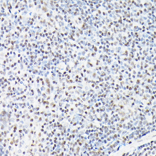
Immunohistochemistry analysis of paraffin-embedded Mouse colon using CTCF Rabbit pAb (CAB13272) at dilution of 1:100 (40x lens). High pressure antigen retrieval performed with 0.01M Citrate buffer (pH 6.0) prior to IHC staining.
Immunohistochemistry analysis of paraffin-embedded Mouse spleen using CTCF Rabbit pAb (CAB13272) at dilution of 1:100 (40x lens). High pressure antigen retrieval performed with 0.01M Citrate buffer (pH 6.0) prior to IHC staining.
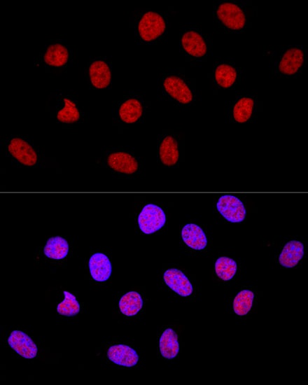
Confocal immunofluorescence analysis of U-2 OS cells using CTCF Rabbit pAb (CAB13272) at dilution of 1:100. Blue: DAPI for nuclear staining.
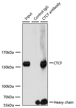
Immunoprecipitation of CTCF from 200 µg extracts of Jurkat cells was performed using 3 µg of CTCF Rabbit pAb (CAB13272). Rabbit Control IgG (AC005) was used to precipitate the Control IgG sample. IP samples were eluted with 1× Laemmli Buffer. The Input lane represents 10% of the total input. Western blot analysis of immunoprecipitates was conducted using CTCF Rabbit pAb (CAB13272) at a dilution of 1:1000.