The CTGF Antibody (CAB11067) is a high-quality antibody developed for reliable detection and analysis of target proteins. The protein encoded by this gene is a mitogen that is secreted by vascular endothelial cells. The encoded protein plays a role in chondrocyte proliferation and differentiation, cell adhesion in many cell types, and is related to platelet-derived growth factor. Certain polymorphisms in this gene have been linked with a higher incidence of systemic sclerosis.
This antibody is validated for use in WB, IF/ICC, ELISA, IF-P applications and has demonstrated reactivity against Human, Rat samples.
Product Name:
CTGF Antibody
SKU:
CAB11067
Size:
100μL, 20μL
Reactivity:
Human, Rat
Conjugate:
Unconjugated
Immunogen:
Synthetic peptide. This information is considered to be commercially sensitive.
Tested Applications:
WBIF/ICCELISAIF-P
Recommended Dilution:
WB
1:1000 - 1:6000
IF/ICC
1:20 - 1:100
IF-P
1:20 - 1:100
ELISA
Recommended starting concentration is 1 μg/mL. Please optimize the concentration based on your specific assay requirements.
The protein encoded by this gene is a mitogen that is secreted by vascular endothelial cells. The encoded protein plays a role in chondrocyte proliferation and differentiation, cell adhesion in many cell types, and is related to platelet-derived growth factor. Certain polymorphisms in this gene have been linked with a higher incidence of systemic sclerosis.
Purification Method
Affinity purification
Gene ID
1490
RRID
AB_2758390
Buffer Information
Store at -20℃. Avoid freeze / thaw cycles. Buffer: PBS containing 50% glycerol, preserved with proclin300 or sodium azide, pH 7.3.
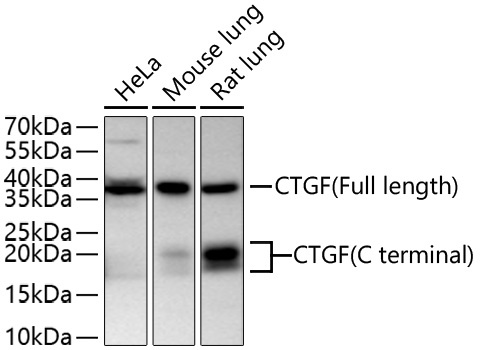
Western blot analysis of various lysates using CTGF Rabbit pAb (CAB11067) at 1:6000 dilution incubated overnight at 4℃. Secondary antibody: HRP-conjugated Goat anti-Rabbit IgG (H+L) (AS014) at 1:10000 dilution. Lysates/proteins: 25 μg per lane. Blocking buffer: 3% nonfat dry milk in TBST. Detection: ECL Basic Kit (AbGn00020). Exposure time: 90 s.
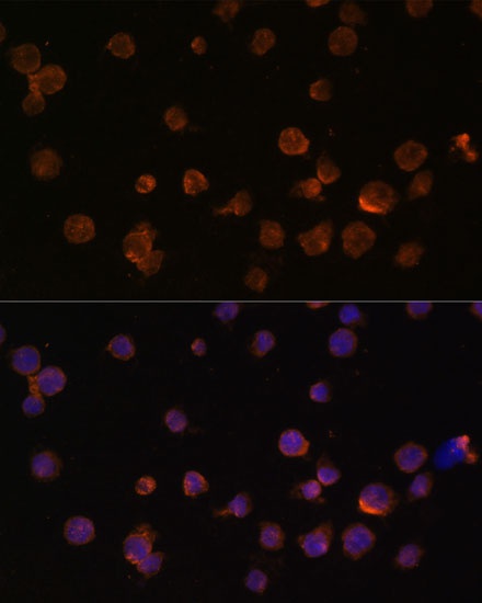
Immunofluorescence analysis of THP-1 cells using CTGF Rabbit pAb (CAB11067) at dilution of 1:100. Secondary antibody: Cy3-conjugated Goat anti-Rabbit IgG (H+L) (AS007) at 1:500 dilution. Blue: DAPI for nuclear staining.